Images reveal best ever sunspot detail
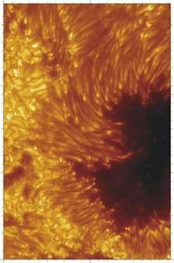
In astronomical imaging, the primary goal is high resolution—the higher the spatial resolution, the more detail that can be seen. With close-up pictures of the sun, hitherto unresolved details of sunspots can help astronomers refine theoretical models. Researchers are now applying a new generation of solar telescopes to that task, obtaining the most highly resolved pictures yet of the Sun's surface (see figure).
Installed in May 2002, the Swedish 1-m Solar Telescope at the Roque de los Muchachos on the Canary island of La Palma recently imaged details as small as 0.12 arcsec (90 km), besting the previous record of 0.2 arcsec (150 km). The evacuated refractor corrects for atmospheric aberrations using a 19-element adaptive optics system, selection of the best images from a continuous real-time stream of exposures, and image restoration using a phase-diversity technique. The adaptive optics in solar observations uses detail from the surface of the sun, such as solar granulation, to correct for daytime "seeing," rather than a nearby star as in nighttime adaptive optics correction.
The images of the penumbra (the edge of a sunspot) reveal never-before-seen dark cores within the bright filaments. The fine-scale structure of the filaments and dark cores changes constantly, lasting more than an hour. The dark cores were found to persist in images of blue and violet wavelengths of 430.5, 436.4, and 487.7 nm. Although the cause of the dark central features of the filaments is not understood, they appear to originate near bright umbral "dots" that stand out near the edge of the dark umbra.
The aperture of the new Swedish 1-m Solar Telescope has a focal length of 20.3 m at a 460-nm wavelength, corrected for chromatic aberrations by an in-vacuum Schupmann corrector. Information from spectrometers and polarimeters at similar resolution is needed to unravel the complex magnetohydrodynamics of such sunspots. The team suggests that further observations of even higher resolution will provide information on fluid velocities and magnetic fields.
REFERENCE
- G. B. Scharmer et al., Nature 420, 151 (2002).
About the Author

Valerie Coffey-Rosich
Contributing Editor
Valerie Coffey-Rosich is a freelance science and technology writer and editor and a contributing editor for Laser Focus World; she previously served as an Associate Technical Editor (2000-2003) and a Senior Technical Editor (2007-2008) for Laser Focus World.
Valerie holds a BS in physics from the University of Nevada, Reno, and an MA in astronomy from Boston University. She specializes in editing and writing about optics, photonics, astronomy, and physics in academic, reference, and business-to-business publications. In addition to Laser Focus World, her work has appeared online and in print for clients such as the American Institute of Physics, American Heritage Dictionary, BioPhotonics, Encyclopedia Britannica, EuroPhotonics, the Optical Society of America, Photonics Focus, Photonics Spectra, Sky & Telescope, and many others. She is based in Palm Springs, California.