Ultrafast pulses promise better processing of fine structures
Ultrafast pulses promise better processing of fine structures
Femtosecond pulses deposit energy into a material before thermal diffusion can occur, making ultrafast sources potentially ideal tools for precision micromachining.
Bruce Craig
During the past several years, advances in more-compact, user-friendly, all solid-state ultrafast laser sources have spurred development of a host of new and exciting applications. One such area is the use of femtosecond and picosecond pulses for micromachining and materials processing. Ultrafast pulses offer the advantage of removing material without significant transfer of energy to the surrounding areas, which can have profound benefits in materials ranging from biological tissue to dielectrics to metals.
One of the fastest-growing segments of the laser market is materials processing. With an average growth rate of 22% over the last five years, the projected market size for 1998 will top the $1 billion mark (see Laser Focus World, Jan. 1998, p. 78, and Feb. 1998, p. 72). For nondiode laser systems, materials processing represents by far the largest market segment. Laser-based materials processing covers a spectrum of applications from heavy industrial welding and cutting to precision processes such as microdrilling and microstructuring. Conventional ultrahigh-power sources--such as CO2 and lamp-pumped Nd:YAG lasers--have dominated the kilowatt-level applications, whereas short-wavelength excimer lasers typically have been the laser of choice for precision machining--that is, structures smaller than 200 µm.
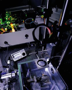
Over the past several years, however, diode-pumped sources have also begun to establish a distinct niche in laser-based precision machining. These sources offer many performance advantages over conventional lasers, such as improved pulse-to-pulse stability, high-quality Gaussian spatial modes, high reliability, more-compact size, and low utility requirements. Several niche applications, such as resistor trimming, memory repair, hard-disk texturing, and rapid prototyping, are now dominated by diode-pumped solid-state (DPSS) sources. And over the next several years, as structure sizes in the microelectronics and microfabrication industries move to even smaller dimensions, both laser sources and processing techniques will continue to evolve to keep pace with the application demands. One area that looks very promising is the use of ultrafast (less than 10 ps) laser pulses for precision machining (see photo).
Advances in ultrafast technology
Until the early 1990s, ultrafast laser technology was traditionally the realm of the "laser jock," so research activities were limited and rather esoteric. With the emergence of Ti:sapphire as a reliable, commercial gain medium with a very wide emission bandwidth, however, laser sources with pulsewidths of less than 20 fs have become commonplace in many research laboratories. The high saturation fluence and the high damage threshold have also allowed Ti:sapphire to become the medium of choice for chirped-pulse amplification systems, the technique by which ultrashort pulses are amplified from a few nanojoules to many millijoules.1
This, coupled with the revolution in diode-pumped green-output lasers (see Laser Focus World, Apr. 1996, p. 63; Nov. 1997, p. 91; and May 1998, p. 123) has meant that high-energy (more than 1 mJ), user-friendly, ultrafast sources--such as the Spectra-Physics/Positive Light Tsunami/Spitfire, the Clark MXR CPA 2000, and the BMI/Coherent Alpha Line--are now widely available to a broad discipline of researchers. Next-generation sources are currently under development in many laboratories, so one can anticipate more ruggedized ultrafast systems becoming available in the not too distant future.
Ultrafast laser material interactions
Ultrafast pulses offer significant potential advantages over conventional sources for micromachining.2 The key benefit of an ultrashort pulse lies in its ability to deposit energy into a material in a very short time period, before thermal diffusion can occur. Following linear or multiphoton absorption of the laser energy, electron temperatures can exceed many thousands of degrees Kelvin. With energy transfer from the electrons to the atomic lattice, material removal, ablation, and plasma formation occur. This competes with thermal relaxation, which is governed by the thermal diffusion length (Dl). This is the distance that the thermal wave can propagate during the laser pulse and is related to the laser pulsewidth tp such that Dl = ktp1/2 , where k is the thermal diffusivity of the material.3
If Dl is shorter than the absorption length, material removal can occur before the onset of energy loss due to diffusion. For nanosecond laser pulses, Dl is typically longer than the absorption length. Ablation occurs from both the melt and vapor phases; as a result the heat-affected zone where melting and resolidification can occur is quite extensive, and the spatial resolution of processed structures is compromised. For ultrashort pulses, on the other hand, the ablation wave actually precedes the thermal relaxation wave so that ablation occurs from the vapor phase. Because the melt phase is minimal, precision hole drilling is possible.
Another benefit of limited thermal diffusion is that the heat-affected zones are significantly reduced. Also, energy loss into the bulk material is minimized, which means that the efficiency of the process is improved and the ablation energy threshold is reduced by, depending on the material, up to two orders of magnitude.
Ultrafast laser metal processing
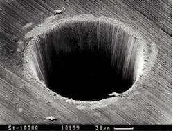
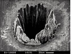
Because of their high thermal conductivity and low melting temperatures, metals pose a particular problem for precision hole drilling. A hole processed with femtosecond pulses is superior to one processed with nanosecond pulses (see Fig. 1).4 For the nanosecond pulse, the thermal relaxation wave propagates into the bulk creating a relatively large layer of melted material, some of which is not removed but resolidifies around the edge of the hole. In addition, the heat-affected zone of the nanosecond hole exhibits a different structure from that of the unprocessed surface. In contrast, the femtosecond hole exhibits sharp edges with no heat-affected zones.
Another benefit of femtosecond pulses is that for an equivalent pulse energy the peak intensity is more than 104 higher than that for the nanosecond pulses, which produces a more efficient ablation process. The laser energy illustrated in Figure 1 is nearly an order of magnitude higher for the nanosecond pulses.
One can speculate about the optimum pulsewidths required to take advantage of the favorable aspects of ultrashort pulse ablation and the minimum features that can be produced. Obviously, this is highly material-specific but the condition that must be satisfied is that material removal occurs in an ablative regime--that is, thermal diffusion is not competitive. Generally, for most materials, this means using pulsewidths shorter than 10 ps--process efficiency typically increases as the pulsewidth gets shorter. A group at the Laser Zentrum Hannover (Hannover, Germany) has demonstrated that, for steel, the critical pulsewidth above which efficiency falls off is about 1 ps.5 With respect to minimum hole size, researchers at the University of Michigan (Ann Arbor, MI) have demonstrated that submicron holes (that is, ten times smaller than the laser spot size) can be generated in silver films by working in a pulse width regime (200 fs) dominated by ablative removal and by taking advantage of the sharp fluence threshold for material removal.6
Real-world applications
Clearly, from a research point of view, the prospect of obtaining beneficial results from machining certain materials with ultrashort pulses looks very encouraging. So what are some of the applications that could induce manufacturers to turn from traditional excimer-laser processing to short-pulse technology?
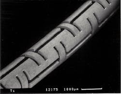
One potential application is production of the metallic and organic stents used as cardiovascular implants (see Fig. 2).5 Using conventional laser sources, the production of stents is limited to only a few (metallic) materials that can be efficiently electropolished (for example, steel) to remove burr and deposited material. Because of the minimally invasive character of ultrashort-pulse processing, a multitude of materials can now be considered for future stents.
Another application is in the automotive industry for production of high-aspect-ratio, high quality micro-holes for fuel injector nozzles (see "Diffractive optics deliver femtosecond pulses to the work-piece," p. 81). A material that is particularly challenging to micromachine is quartz, which is widely used in optoelectronics, micro-optics, and fiber technology. At pulsewidths of less than 200 fs, highly reproducible channels of less than 20-µm diameter can be generated over lengths of greater than 1 mm in quartz substrates.7
Energetic materials--because of their sensitivity to shock or thermal stress--also benefit from ultrashort-pulse micromachining. The possibility of processing these materials with ultrashort pulses and avoiding unintentional initiation has been demonstrated--researchers at Lawrence Livermore National Laboratory (LLNL; Livermore, CA) showed that there is a dramatic difference in cutting PETN explosives with 150-fs versus 500-ps pulses.8 Analysis of walls cut with femtosecond pulses shows that they are essentially unchanged by the machining process.
Ultrafast pulses in medicine
A somewhat different area of laser materials processing is medicine, in which lasers are used for a variety of surgical procedures. Several groups have recognized that the many potential benefits of ultrashort-pulse micromachining also apply to hard-tissue ablation. One of the major concerns of using conventional lasers to ablate calcified tissue is that fractures and fissures can accompany laser treatment because hard tissue is prone to shear and compressive stresses. Ultrashort-pulse tissue ablation causes minimal collateral mechanical and thermal damage because of the short energy deposition time and the high efficiency of the ablation process--most of the deposited energy is removed in the ablated material. The fact that minimal energy is deposited into the surrounding tissue also means less patient discomfort.
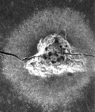
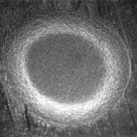
With this in mind, researchers at LLNL have explored the use of ultrashort pulses for dentistry applications.9 Long-pulse ablation of a tooth causes the tooth to crack and produces a random-shaped hole with collateral damage. In contrast, ultrashort-pulse laser drilling produces a clean hole with no thermal damage (see Fig. 3). Whether or not a high-energy ultrafast laser drill could provide dentists with a cost-effective alternative to the current mechanical drill, however, remains to be seen.
Another benefit of ultrashort pulses for surgical procedures is the inherent precise spatial control--both in terms of location and depth of penetration. This could be useful for very sensitive procedures such as spinal surgery where collateral damage can have devastating consequences (see "Ultrafast lasers pin down tissue types," p. 86). Ultrashort-pulse procedures have also been explored for transmyocardial revascularization, a minimally invasive procedure in which small holes are drilled through the heart muscle (myocardium) into the inner chambers.10 Such channels appear to improve blood flow to the myocardium, which improves a patient`s exercise tolerance and provides relief from anginal pain. Currently the procedure is accomplished with excimer or CO2 lasers.
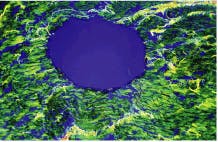
A comparison of holes drilled in pig myocardium with an excimer laser and an ultrashort-pulse laser shows extensive thermal damage in the excimer-treated tissue, whereas the hole generated with ultrashort pulses is free of thermal damage (see Fig. 4). The advantages of reduced collateral damage with ultrashort pulses could have significant implications for this application. Other areas that also may benefit from ultrashort-pulse laser ablation are certain ophthalmic procedures--including glaucoma treatment, cataract removal, and laser keratomileusis.11
Looking ahead
Clearly ultrafast laser pulses show great potential for processing fine structures in a variety of materials. Will we expect to see ultrafast sources in the operating room or production floor in the near future? Before this can happen, ultrafast sources need to continue their development from today`s R&D systems to hands-off, ruggedized, reliable, compact devices with a simple user interface. All-solid-state diode-pumped laser architecture is key to achieving these goals. At Spectra-Physics we took a step in this direction in May when we released at CLEO `98 (San Francisco, CA) the first commercial, all-solid-state diode-pumped Ti:sapphire amplifier system.
Ultrafast sources also will need to compete with improvements in micromachining techniques that can be anticipated from conventional laser sources. Ultimately it will come down to the economics of the process--throughput, yield, quality, cost, reliability, and such--and whether ultrafast laser-processed materials really offer tangible benefits over competitive techniques. In regard to microdrilling and microstructuring at dimensions of less than 100 µm, today`s competing technologies are electrodischarge machining (EDM) and focused ion beam (FIB) machining. Both processes are equipment-intensive and have limitations, so the prospects for ultrafast-pulse alternatives look promising. And with the growth of the electronics, medical, and environmental-sensing markets and the push to smaller-sized structures, development of the laser-based micromachining industry is assured well into the next millennium. o
ACKNOWLEDGMENTS
The author wishes to thank Stefan Nolte of the Laser Zentrum Hannover and John Marion of LLNL for their assistance in creating this article.
REFERENCES
1. D. Strickland and G. Mourou, Opt. Comm. 56, 219 (1985).
2. X. Liu and G. Mourou, Laser Focus World, Aug. 1997, p. 101.
3. E. Matthias et al., Appl. Phys. A. 58, 129 (1994).
4. C. Momma et al., Opt. Comm. 129, 134 (1996).
5. S. Nolte et al., CFD3, CLEO 1998 Abstracts, p.510.
6. P. P. Pronko et al., Opt. Comm. 114, 106 (1995).
7. H. Varel et al., Appl. Phys. A. 65, 367 (1997).
8. P. S. Banks et al., CFD2, CLEO 1998 Abstracts, p. 510.
9. Joseph Neev et al., IEEE J. of Select Topics in Quantum Elect. 2, 790 (1996).
10. J. Marion, "Ultrashort pulse laser surgery," LLNL, UCRL-MI-12940 (1997); see also http://lasers.llnl.gov/lasers/mtp)
11. See M. Ito et al., J. Refractive Surg. 12, 721 (1996) and R. M. Kurtz et al., Proc. SPIE Vol. 3255, in press.
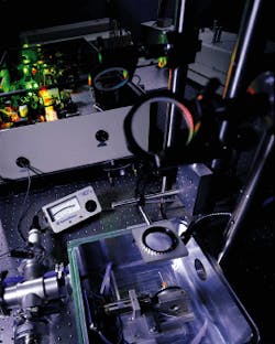
Ultrashort pulses from a Tsunami Ti:sapphire laser amplified by a Spitfire regenerative amplifier are routed through three mirrors and a focusing lens into an aluminum vacuum chamber (shown here opened for adjustment) to machine a small steel tube to be used as a stent in cardiac surgery.
FIGURE 1. Hole drilled with ultrafast pulses (pulse duration of 200 fs, at 120 µJ, delivered 0.5 J/ cm2) in 100-µm-thick steel foil (top) is superior to hole processed with nanosecond pulses (pulse duration of 3.3 ns, at 1 mJ, delivered 4.2 J/cm2 (bottom). In each case, the minimum laser fluence was used.
FIGURE 2. Titanium stent (medical implant) was machined with a femto second laser.
FIGURE 3. Teeth drilled with nanosecond pulses exhibit local heating and cracking from the large thermal stresses (top); femtosecond pulses produce a clean hole with no collateral damage (bottom).
FIGURE 4. Histological sections of an excimer-laser-drilled pig myocardium exhibit extensive thermal damage (dark areas) to the tissue surrounding the hole (top); ultrafast laser produces a smooth-sided hole free of thermal damage to surrounding tissue (bottom).
Diffractive optics deliver femtosecond pulses to the work-piece
One of the major problems to be overcome if ultrashort pulses are to be used in micromachining applications is actually delivering the beam to the work-piece. When conventional imaging and beam focusing geometries are used, the beam intensity in the focal plane is so high that ionization, plasma formation, and other nonlinear effects--such as self-phase modulation, self-focusing, and filamentation--occur in the surrounding air. This results in significant distortion of the beam spatial profile and requires, therefore, that the processed parts be placed in a vacuum. Many of the examples discussed in the main article were processed in a vacuum. Clearly, such a requirement poses a serious impediment for widespread acceptance of ultrafast micromachining technology.
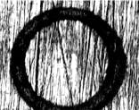
At the Laser Zentrum Hannover (Hannover, Germany), we have demonstrated that a diffractive optical element (DOE) can minimize nonlinear beam distortions and produce high-quality holes in metallic work-pieces at atmospheric pressure.1 We generated a donut-like intensity pattern and maintained the B integral at less than 0.14 using a DOE that is a combination of a Fresnel lens and a phase plate (see left image). The B integral is a quantity governing spatial and temporal distortions of the beam due to nonlinear effects--to avoid such effects B should generally be less than 1.
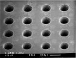
The other benefit of a DOE is that energy throughput is nearly twice as efficient as that of a typical imaging system. A DOE system has been used with 1.3-mJ, 150-fs pulses for production of high-aspect-ratio, burr-free holes in steel that are required for fuel injection nozzles in the automotive industry. The machining was done in air and the process was highly reproducible (see right image).
Stefan Nolte, Carsten Momma, and Günter Kamlage
STEFAN NOLTE, CARSTEN MOMMA, and GÜNTER KAMLAGE are staff scientists at the Laser Zentrum Hannover, Hannover, Germany.
REFERENCE
1. C. Momma et al., Appl. Phys. A, "Beam delivery of femtosecond laser radiation by diffractive optical elements," in press.
Donut-shaped intensity pattern (left) was produced in copper with the beam delivered via a diffractive optical element system, which was also used to produce an array of holes in steel (right). The holes were drilled in air with 1.3-mJ, 150-fs pulses.
Ultrafast lasers pin down tissue types
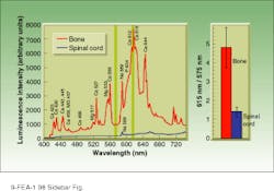
To improve the usefulness of ultrafast lasers in biomedical applications, researchers at Lawrence Livermore National Laboratory (LLNL; Livermore, CA) have been developing diagnostics that can discriminate between different tissue types. Our system uses the luminescence created by the plasma formed during tissue ablation to discriminate between bone and soft tissue. The spectral characteristics of the plasma depend on the material that was heated to form the plasma--each element has a distinct signature (see figure).
Bone has significant mineral lines, whereas nerve tissue has few distinctive spectral characteristics. By analyzing the spectral luminescence signatures of different tissue types we have developed a system that can control the laser ablation so that only the desired tissue is ablated. Our diagnostic system senses whether the laser is cutting bone or nerve tissue and shuts the laser off if it is beginning to cut nerve. The luminescence signal is collected by a fiberoptic near the focal point of the ultrafast laser. Fast electronics measure the ratio of two wavelengths, one a strong calcium peak at 616 nm and the second at 570 nm, an area where there is little luminescence signal.
While ablating bone, the ratio of these signals is about 5, whereas when nerve is ablated, the ratio drops to about 1.2. Operating at a pulse repetition rate of 1 kHz, we can block the laser output (by deactivating one Q-switch) before the next laser pulse if the ratio of the luminescence signals drops below 2. In this way our system instantly determines whether the laser is cutting bone (high calcium signal) or nerve (low calcium signal). When the calcium signal drops, the laser shuts off, preventing the surgeon from accidentally cutting nerve.
John Marion and Beop-Min Kim
JOHN MARION and BEOP-MIN KIM are at Lawrence Livermore National Laboratory.
Prominent calcium peaks in the luminescence signals for bone (red) and spinal cord (blue) allow a surgeon to distinguish between bone and spinal cord or other soft tissue. Locking out the laser from ablation of nerve and soft tissue prevents damage to surrounding areas during bone removal.
