QCL technology poised to transform IR spectroscopy, microscopy
Spectral imaging is an extremely powerful analytical tool that combines chemical and morphological information to identify and quantify the distribution of molecular species. A wide range of fields, including biology, forensics, and materials science, regularly use infrared (IR) microscopes to generate label-free spectral images. The IR microscope’s origin can be traced back all the way to 1949, when a reflection-based microscope was fitted with a dispersive single-beam IR spectrometer.1 For much of the next 30 years, IR microscopes were little more than a scientific novelty. It was not until 1977 when scientists coupled a Fourier-transform infrared (FTIR) spectrometer to a microscope2 that they started producing analytical-quality data.
Over the proceeding decades, FTIR microscopy quickly became the go-to method for microchemical analysis. While there have been many incremental changes to the technology, until recently there has been little change to the foundational methodology behind IR microscopes.
Fundamentals of IR microscopy design
Most FTIR microscopes are designed to work in the mid-infrared (mid-IR) region of the spectrum, from 2.5 to 25 μm (4000 to 400 cm-1) because it directly corresponds to the fundamental vibrational frequencies of covalently bound molecules. In many ways, this makes mid-IR absorption spectroscopy the simplest molecular spectroscopy since it is a direct measurement technique. At the same time, the mid-IR region has significant optical challenges because the wavelength range is so vast, and the photon energies are so low.
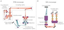
The traditional FTIR approach uses an off-axis parabolic mirror to collimate a broadband thermal emission source, called a globar, and send it through a scanning interferometer. The sample leg of the interferometer incorporates a reflective condenser to illuminate the sample and a Schwarzschild (reflective) objective to recollimate the transmitted light. Finally, the light is measured with an IR photodetector, and Fourier transformed to reconstruct the transmission spectrum (see Fig. 1). Despite all of this complexity, the robust nature of this technique still fueled the commercial success of FTIR microscopy through the early 2000s. However, as other microspectroscopy techniques, such as confocal Raman, became more readily available, the comparative performance of IR microscopy started to wane.
Now, it appears IR microscopy is amid yet another groundbreaking revolution due to the commercial availability of QCLs between 3 and 13 μm (3300–770 cm-1). Not only are modern QCLs tunable over a frequency range of several hundred wave numbers with linewidths <1 cm-1, they are also inherently single-spatial-mode and linearly polarized. As a result, replacing the globar with a bank of QCLs (typically four) improves the optical performance of the microscope and eliminates the need for an interferometer altogether. Therefore, leading to the development of an entirely new form of IR microscopy, often referred to as discrete frequency infrared (DFIR) or laser direct infrared (LDIR) microscopy. Even though this technology is relatively new, there are already a handful of commercially available QCL-based microscopes from companies such as DRS Daylight Solutions (San Diego, CA) and Agilent (Santa Clara, CA).In April 2021, Dr. Rohit Bhargava, an expert in IR microscopy at the University of Illinois at Urbana-Champaign (Urbana, IL), along with Yamuna Phal (a graduate student) and Kevin Yeh (a postdoctoral candidate), published an extremely detailed review of QCL-enabled IR microscopes.4 In this article, they discussed technical considerations that should be accounted for when choosing the appropriate laser, detector, and optics for a DFIR microscope.
The QCL imaging advantage
Perhaps the single most significant advantage of using QCLs instead of broadband light sources for IR microscopy is spectral irradiance at the sample. Even though typical silicon nitride globars produce several watts of optical power, it is spread out over a relatively large solid angle and an even larger spectral range. By contrast, typical QCLs produce hundreds of milliwatts of average power within a few-milliradian divergence angle. Therefore, it is no wonder that QCL-based IR microscopes can acquire data exponentially faster than traditional FTIR microscopes. In a typical DFIR system, the limiting factor on sample acquisition is set by the damage threshold of the sample.
One striking example of the difference between QCL and FTIR microscopes was demonstrated by Claus Kuepper, Angela Kallenbach-Thieltges, and others at Ruhr University Bochum (Bochum, Germany)—they evaluated the use of QCL-based IR microscopy as a means of cancer diagnostics.5 In their study of 110 patients with colorectal cancer, published in Nature’s Scientific Reports, they managed to show 96% sensitivity and 100% specificity compared to histopathology. But according to the authors, “The main hurdle for the clinical translation of IR imaging is overcome now by the short acquisition time for high-quality diagnostic images.”
In the supplemental material,6 they showed a comparison of a gold standard hematoxylin and eosin stain (see Fig. 2a), an image taken with a Spero QT QCL-based IR microscope from DRS Daylight Solutions (see Fig. 2b), and a FTIR microscope (see Fig. 2c). Both images were able to identify tumorous regions and infiltrating inflammatory cells; what is important to point out is that the QCL-based system was able to measure the sample in 34 minutes. In contrast, the FTIR image took ~5400 minutes.In a 2017 article published in Analyst, Bhargava’s group showed similar results when studying polymer samples using both FTIR- and QCL-based IR microscopes.7 Using a prototype QCL-based microscope from Agilent, they measured polyethylene glycol (PEG) films using both s- and p-polarized light to determine the macromolecular orientation successfully. For comparison, the FTIR system took 1620 minutes to collect the spectral image, whereas the QCL-based system took a mere 9 minutes.
Looking to the future
Bhargava, Phal, and Yeh summarized it best in the conclusion of their recent review article: “...technology has pushed IR spectroscopic imaging beyond previously foreseeable limits.” While there are still significant cost hurdles associated with integrating a bank of four QCLs into an IR microscope, the advantages for IR spectral imaging are clear. As QCL production continues to improve over time, there is no doubt prices will continue to trend down as capabilities trend up.
ACKNOWLEDGMENT
The author would like to thank Ellen Miseo, Chief Scientist at TeakOrigin and current President of the Coblentz Society, for her input on the current state of QCL-based IR microscopy.
REFERENCES
1. R. Barer, A. Cole, and H. W. Thompson, Nature, 163, 4136, 198–201 (1949).
2. R. Cournoyer, J. C. Shearer, and D. H. Anderson, Anal. Chem., 49, 14, 2275–2277 (1977).
3. See http://bit.ly/IRMicrospectroscopy.
4. Y. D. Phal, K. Yeh, and R. Bhargava, Appl. Spectrosc., 00037028211013372 (2021).
5. C. Kuepper et al., Sci. Rep., 8, 1, 1–10 (2018).
6. C. Kuepper et al., Sci. Rep., 8, 1, supplement (2018).
7. T. P. Wrobel, P. Mukherjee, and R. Bhargava, Analyst, 142, 1, 75–79 (2017).
About the Author
Robert V. Chimenti
Director, RVC Photonics LLC
Robert V. Chimenti is the Director of RVC Photonics LLC (Pitman, NJ), as well as a Visiting Assistant Professor in the Department of Physics and Astronomy at Rowan University (Glassboro, NJ). He has earned undergraduate degrees in physics, photonics, and business administration, as well as an M.S. in Electro-Optics from the University of Dayton. Over a nearly 20-year career in optics and photonics, he has primarily focused on the development of new laser and spectroscopy applications, with a heavy emphasis on vibrational spectroscopy. He is also very heavily involved in the Federation of Analytical Chemistry and Spectroscopy Societies (FACSS), where he has served for several years as the Workshops Chair for the annual SciX conference and will be taking over as General Chair for the 2021 SciX conference.

![FIGURE 2. Comparison of the IR-based tissue analysis. Colorectal cancer tissue analysis using hematoxylin and eosin stain (a), QCL-based microscope (b), and FTIR-based microscope (c). [6] FIGURE 2. Comparison of the IR-based tissue analysis. Colorectal cancer tissue analysis using hematoxylin and eosin stain (a), QCL-based microscope (b), and FTIR-based microscope (c). [6]](https://img.laserfocusworld.com/files/base/ebm/lfw/image/2021/07/2107LFW_rob_2.60e72012c0e91.png?auto=format,compress&fit=max&q=45?w=250&width=250)
