MEMS swept-wavelength laser enables new OCT capabilities
Optical interferometry is sensitive and noninvasive, making optical coherence tomography (OCT) a valuable clinical diagnostic tool. OCT directs light to a target tissue, detects the reflection, and processes the reflection to reconstruct an image of subsurface structures. Although powerful, long acquisition times make some endoscopic imaging either impractical or impossible.
The key to enabling new applications is to increase the A-scan speed—the acquisition speed of the depth profile at a single location. In swept-source OCT (SS-OCT), a light beam sweeps through a range of wavelengths and the reflected signal is processed to recover a depth profile at that point. The point is scanned over an area of interest to generate a 3D representation of subsurface tissue. The speed of SS-OCT is limited by the wavelength sweep rate. Massachusetts Institute of Technology (MIT; Cambridge, MA) researchers led a multidisciplinary development effort incorporating a rapidly sweeping laser source into a proof-of-principle multi-megahertz SS-OCT system, demonstrating new clinical imaging capabilities and opening the pathway for low-cost turnkey commercial systems.
Wavelength scanning: Just the start
Under a National Institutes of Health Small Business Innovation and Research grant, Praevium Research (Santa Barbara, CA) has developed a microelectromechanical vertical-cavity surface-emitting laser (MEMS-VCSEL) consisting of a MEMS cavity mirror suspended on struts above a wide-gain-bandwidth optically pumped active region. The suspended mirror is electrostatically driven, with restoring force supplied by the struts. The device was engineered with struts stiff enough to support oscillation frequencies up to 1.5 MHz. The mirror motion modifies the resonant cavity, leading to a wavelength sweep over a range of more than 100 nm.
In SS-OCT, the laser beam is split, with part directed to and reflected from wherever there is a change in index of refraction in the target tissue, such as at cell and tissue boundaries. The instantaneous returned signal is the superposition of signals reflected from different depths. The other portion of the beam traverses a reference arm. When the reflected and reference beam light recombine, the resulting intensity is the sum of all the constructively or destructively interfering signals. As the wavelength is scanned, the net intensity varies, corresponding to the constructive and destructive interference at that wavelength to form an interferogram signal.
In the MIT demonstration, the interferogram signal is collected at a fixed sampling rate. If the wavelength sweep were perfectly linear in wavenumber with respect to time, the signal could be converted to an intensity vs. depth profile, called an A-scan, through a Fourier transform. In practice, small deviations from linearity in the sweep become significant, especially at high data rates, such as the 2 gigasamples per second used to provide the required imaging range for this application. So a measurement of wavenumber, k, with respect to time is required to calibrate the signal before the Fourier transform.
Here, that measurement is implemented with an integral Mach-Zehnder interferometer (MZI) whose signal is captured simultaneously with the OCT data. It’s computationally intensive, but according to Ben Potsaid, a research scientist at Thorlabs (Newton, NJ) and a visiting scientist at MIT, “advances in graphics processing unit (GPU) accelerated numerical processing techniques are making real-time processing of the incoming data stream possible at a modest budget.”
As in any engineering system, tradeoffs must be made, and changes in one part of a system force changes in other parts. Here, faster wavelength sweeping repetition rates with the MEMS-VCSEL forced the adoption of an integral MZI with dual channel acquisition, and that additional data forced the incorporation of GPU processing. The high acquisition speed also required higher power output, supplied by a custom-designed Thorlabs booster optical amplifier.
Faster A-scans, more extensive OCT capabilities
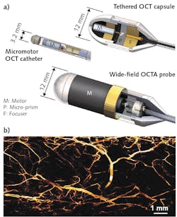
For this demonstration, the research team adopted OCT scan rates of 2.4 and 3 MHz in conjunction with three different optical probes. “Faster imaging rates make it possible to image structures where the long acquisition time was previously impractical,” Potsaid says. For example, OCT angiography is challenging, requiring rapid, repeated scanning to detect blood vessel flow patterns that may be connected with disease. That probe uses a stable brushless, ball-bearing DC motor to rotate a prism at 600 Hz. This generates, for example, full circumferential scans of blood vessels in the swine lower GI tract with axial resolution of about 12 µm and transverse resolution of 30 µm, to a depth of approximately 1.5 mm.
“Our initial results,” says Jason Zhang, a doctoral student at MIT, “establish the feasibility of multi-megahertz OCT and OCT angiography with a pathway toward future small and low-cost OCT systems based on MEMS-VCSEL technology.”
About the Author

Richard Gaughan
Contributing Writer, BioOptics World
Richard Gaughan is the Owner of Mountain Optical Systems and a contributing writer for BioOptics World.