Laser brazing dissimilar metal medical devices
Seamless joining eliminates adhesives
Gen Satoh and Y. Lawrence Yao
The joining of dissimilar materials is a critical issue in the continued development of advanced medical devices due to the unique properties possessed by materials such as nickel titanium (NiTi), platinum (Pt), and stainless steel (SS). Requirements for joining dissimilar biocompatible materials can stem from introducing unique functionalities through the use of shape memory alloys such as NiTi in conjunction with SS, or for decreasing costs while maintaining exceptional corrosion resistance in Pt-to-SS joints. To address these requirements, a new laser joining process is under development to form autogenous (no filler material) joints between dissimilar, biocompatible metal pairs. The autogenous laser joining process would enable seamless joining of these components and eliminate the need for proprietary adhesives and filler materials used in many current designs.
Laser-based joining processes, due to their low thermal input and small spot size, have become the primary joining mechanism of metallic parts in medical devices such as pacemakers. The advantages of lasers in joining processes over conventional heat sources, such as minimal heat-affected zones and controlled energy delivery, are vital to medical device manufacturing processes due to the thermal sensitivity of components as well as their continued miniaturization. Dissimilar metal joints, however, are often complicated by the formation of new phases such as brittle intermetallics within the joint that leads to low strength and premature failure.
Autogenous laser brazing
While the majority of laser-based joining processes use the laser input to directly melt the base or filler materials at the joint, the autogenous laser joining process is designed to form joints that are significantly smaller than the laser beam spot size by taking advantage of heat accumulation at the joint interface. This joining process involves laser irradiation of one of the base materials which is scanned toward the dissimilar metal interface.
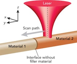
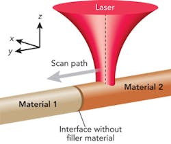
Laser parameters such as power and speed are chosen such that the equilibrium temperature of the irradiated piece does not exceed its melting temperature. Heat accumulation due to the thermal resistance of the interface causes the temperature to rise above the melting temperature of one of the base materials as the laser beam approaches, forming a molten layer. The laser beam is turned off as it reaches the interface and the melt layer is quenched when it comes in contact with the adjacent cold work piece, forming an autogenous braze-like joint. A schematic diagram of the joining process is shown in FIGURE 1.
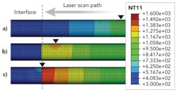
The autogenous laser brazing process aims to minimize mixing of the two materials due to the small melt volume, high quench rate, and localized melting of one side of the weld joint to reduce brittle intermetallic formation. Another benefit of the localization of heat is that it allows for joining of material pairs with similar melt temperatures with minimal mixing. FIGURE 2 shows temperature profiles from a thermal model of a laser beam scanning toward a wire-to-wire interface. An increase in temperature as the laser beam reaches the interface is observed due to heat accumulation.
Experimental setup
NiTi and stainless steel 316L wires, roughly 380 µm and 368 µm in diameter, respectively, were chosen for joining experiments. NiTi and SS have received particular attention for use in medical devices; stainless steel for its low cost and proven biocompatibility, and NiTi for its super elastic and shape memory properties. Prior to joining, one end of each wire was ground flat, perpendicular to the axis of the wire, and assembled in a welding fixture. The ground faces of the samples were held together using an axial force during irradiation by a continuous-wave Nd:YAG laser operating at a wavelength of 1064 nm. The Gaussian laser spot was controlled to be the same size as the diameter of the wires. Laser power was adjusted up to a maximum of 4.75 W while scan speed and scan distance were adjusted between 0.2–1 mm/s and 1–2.5 mm, respectively. Laser joining was performed in an inert environment of ultrahigh-purity argon gas which was flowed into the welding fixture.
Weld geometry
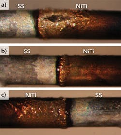
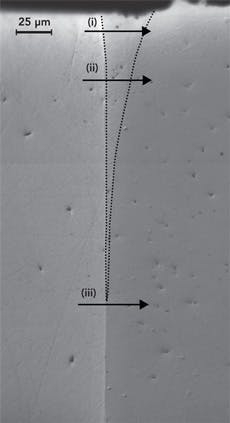
FIGURE 3 shows a typical joint created using the autogenous laser brazing process. Overall, the joint shows a clean outward appearance with no obvious signs of porosity or cracking. No large-scale deformation of the wire is observed, indicating that significant melting of the base materials did not occur during processing. FIGURE 4 is an optical micrograph of the same sample after cross-sectioning along the Y-Z plane. A clean interface is observed with little to no porosity, no cracking, and no signs of incomplete joining at the interface. The joint itself is narrowest toward the center of the wires and wider along the top and bottom surfaces.
Compositional analysis
Quantitative energy-dispersive x-ray spectroscopy (EDS) profiles performed across the NiTi-SS interface at different depths from the irradiated surface are shown in FIGURES 5a–5c. Between the two base metal compositions is the joined region showing a mixture between Fe, Cr, Ni, and Ti. The extent of this mixed region indicates the width of the joint. This specific sample shows joint widths ranging from between roughly 5–25 µm. The different shapes of the composition profiles suggest that different joining mechanisms are dominant along different regions of the joint. Toward the laser-irradiated surface the melted layer thickness is greater, which indicates a longer melt duration, allowing greater dilution of the stainless steel into the molten NiTi. Toward the center of the wires, where minimal mixing of the two materials is observed, the melt layer thickness was likely significantly smaller. The composition profile in this region resembles a diffusion-controlled process while the upper layers resemble a fusion-based joining mechanism. FIGURE 5 also shows that the joint width is significantly smaller than the beam spot size of 400 µm — this confirms that the mechanism of joint formation is not direct melting by the laser beam, but thermal accumulation as desired.
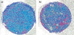
FIGURES 6a and 6b show compositional maps over the fracture surfaces of a joint created using the autogenous laser brazing process, where red, green, and blue represent Fe, Ni, and Ti, respectively. Both fracture surfaces are dominated by green and blue, which indicates that fracture occurred in the Ni- and Ti-rich regions of the joint. Quantitative measurement of the composition puts the fracture surface within the joint rather than within one of the base materials. Regions with primarily red coloring indicate incomplete joining or fracture in the base SS wire. The existence of incomplete joining at the interface was observed to be a function of scan speed at constant power. As the laser scan speed is increased, the overall energy input into the wire is reduced. This can have the effect of decreasing the melt layer thickness or eliminating the melt layer all together, causing insufficient melting to cover the entire faying surfaces.
Joint strength
An ideal joint between dissimilar materials will have a strength that exceeds the weaker of the base materials. The stainless steel wire yields at an applied load of roughly 280 MPa and fractures at a load of nearly 560 MPa after significant plastic deformation. The super elastic NiTi sample initially deforms elastically until the phase transformation from austenite to martensite occurs at a stress level of 470 MPa. After the load plateau, another linear elastic region is observed, and fracture is observed at nearly 1.5 GPa of applied stress.
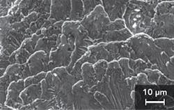
FIGURE 7 shows a typical fracture surface as seen through electron microscopy for a sample joined using the autogenous laser joining process. The surface has a morphology indicative of quasi-cleavage fracture, and as discussed above, fracture is believe to be occurring within the joint, not in the base material. For experiments at low scan speeds (up to 1 mm/s), the maximum joint strength reaches roughly 275 MPa. While this strength is roughly equal to the yield stress of stainless steel, appreciable plastic deformation within the stainless wire before fracture is not observed. Recent experiments at higher velocities and powers (up to 2 mm/s at 8 W) have shown fracture stresses exceeding 470 MPa. It is believed that the increased scan speed allows for greater heat localization and accumulation to form a smaller melt pool over a shorter time duration, leading to reduced mixing between base materials and reduced intermetallic formation.
Conclusion
A new joining process, laser autogenous brazing, has been investigated for creating seamless joints between two biocompatible materials, NiTi and stainless steel 316L, for medical device applications. The joints show strengths that approach the yield stress of the stainless steel base material during tensile testing and have fracture surfaces indicative of quasi-cleavage fracture.
Recent experimental results show further increases in fracture strength to levels above the yield stress of the stainless steel. EDS mapping of the cross-sections indicated that joint widths were significantly smaller than the incident laser beam diameter, suggesting melt pool formation due to heat accumulation. The joint strengths achieved suggest that laser autogenous brazing is a viable and promising method for creating robust joints between the NiTi and stainless steel biocompatible dissimilar material pair without filler materials, and is expected to be applicable to a wide variety of other dissimilar metal pairs. ✺
Gen Satoh and Y. Lawrence Yao are with the Manufacturing Research Lab, Columbia University, New York, NY, [email protected].
More Industrial Laser Solutions Current Issue Articles
More Industrial Laser Solutions Archives Issue Articles
