Optical diagnostics continue migration from benchtop to bedside
Will optical tools ever find their footing in mainstream medicine? With more laser companies focusing on the biomedical market, the realities of bringing these products to market are receiving some serious attention.
The notion of an optical tool that can achieve early, accurate, and—perhaps most important—noninvasive detection and diagnosis of disease has intrigued technologists and physicians (not to mention patients) for years. The ability to build laser-based diagnostic devices that are compact, low-cost, user friendly, and deliver all that we expect them to, however, has been anything but easy.
At some points the difficulty has rested with the technology, at others, the audience. In recent years, it has been the enormous cost involved in bringing optically based medical products to market, especially in the United States. According to those at the forefront of biomedical optics research and development, however, the landscape is changing. The result should be a market finally coming to fruition after years of frustration and fine-tuning.
"The last few years have seen a damper on private-sector investment in biomed," said Irving Bigio, professor of biomedical, electrical, and computer engineering at Boston University (BU; Boston, MA). "But this is changing."
In 2003, for example, the National Institutes of Health (NIH) began funding three multicenter clinical translational studies in biomedical optics. Given that these studies are designed to determine both clinical viability and commercial possibilities, this is a major shift in U.S. funding of medical technology development. Among those benefiting from this new approach is the Beckman Laser Institute (Irvine, CA), which received a $7 million grant from the National Cancer Institute (NCI; part of the NIH) to speed the development of diffuse optical imaging for the detection and analysis of tumors. The goal is to create commercial laser-imaging devices that complement conventional cancer-detection methods.
"There are many opportunities for translational research in biomedical optics to take this technology from the benchtop to the bedside," said Bruce Tromberg, director of the Beckman Laser Institute. "People in this field are now capitalizing on the insights gained in basic studies of tissue and light interaction by engineering systems that can acquire disease-specific information and validate the technology in the clinical setting."
As with many of the potential markets for lasers, optical diagnostics has suffered in part from the old adage "a technology in search of an application." Thus not only is this new wave of government support important, so is the need for R&D groups to make product-development decisions based as much on the potential markets and applications for these devices as on the capabilities of the technology.
"The sad fact is that most academics do not have a clue about manufacturing issues," said Warren Grundfest, M.D., chairman of the Biomedical Engineering Program at University of California-Los Angeles. "It is not just 'can the technology do something nothing else can do,' but can it do it cost-effectively. Company after company (coming out of telecom) has called me looking for a cheap, quick solution (in biomed), and I give them all the same advice: if you think this is going to be quick and easy and only cost you $1 million, you are delusional."
Some success
Despite the technological and market hurdles, however, optical diagnostics has not been without commercial success. Several optical technologies are being used by ophthalmologists for retinal diagnostics, for example; Imaging Diagnostic Technologies (San Diego, CA) claims to have sold more than 2500 of its GDx Scanning Laser Polarimeters, which use an IR laser for glaucoma detection.
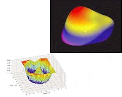
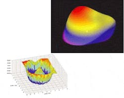
Similarly, wavefront analysis has found a golden niche in laser vision-correction surgery. Wavefront analyzers, such as the CustomVue from VISX (Santa Clara, CA), precisely measure the overall refractive error of the entire eye, including any aberrations caused by the tear film, anterior and posterior cornea, lens, vitreous, and retina (see Fig. 1). More-standard approaches such as topography can define corneal irregularities but cannot detect aberrations in other parts of the eye.
Another high-profile success story in optical diagnostics is optical coherence tomography (OCT). Invented by James Fujimoto, Eric Swanson, and colleagues at the Massachusetts Institute of Technology (MIT; Cambridge, MA), OCT is an optical technique that obtains high-resolution images of tissue by focusing a beam of near-infrared light into the tissue and measuring the intensity and position of the resulting reflections. The MIT technology was originally licensed to Humphrey Instruments (now Humphrey Zeiss, a subsidiary of Carl Zeiss Meditec) for ophthalmic diagnostic applications; Humphrey introduced the first commercial OCT product in 1996, and today the third generation of this system, the Stratus OCT3, is used to diagnose macular degeneration and other retinal conditions such as glaucoma and diabetic retinopathy.
"In OCT, 830 nm turns out to be optimum for imaging the retina, while 1300 nm turns out to be optimal for imaging pretty much everywhere else, such as arteries and the GI tract," said Joseph Izatt, associate professor of biomedical engineering at Duke University (Durham, NC) and a leading researcher in biomedical optics, spectroscopy, and optical and ultrasonic imaging.
In fact, OCT is being investigated for several topical and endoscopic applications in gastroenterology, dermatology, dentistry, cardiology, and other medical disciplines, particularly for early cancer detection. Other potential applications include vascular imaging and molecular biology (see Fig. 2).
FIGURE 2. Researchers at Duke University are using 3-D optical coherence tomography to study chick embryo hearts and gain more insight into the process of cardiovascular development. A 3-D reconstruction of the outflow limb and presumptive right ventricle with a cutaway of the outflow limb and the single ventricle reveals internal structures of the early heart tube (left). A 3-D reconstruction of a chick heart at three days old is shown at right. Still images of the same hearts show cross-sectional OCT images in the sagittal plane (inserts). CJ indicates cardiac jelly; en, endocardium; IC, inner curvature; i, inflow limb; m, myocardium; o, outflow limb; and v, presumptive ventricle.1
"Optical coherence tomography is great for small-animal imaging and noninvasive imaging for research into functional genomics and drug discovery," Izatt said. "Three-dimensional noninvasive imaging of small-animal organs is a very competitive area. The problem with techniques such as micro-MRI is that they are expensive and time-consuming. You can get almost the same resolution (as OCT), but you have to put the animal in the MRI machine for 20 hours."
Further out
Despite these and other advances, and even with the shift to more applied and functional R&D projects, most optically based diagnostic tools are still years away from mass commercialization. Fluorescence and Raman spectroscopy, for instance, are well-established techniques involving fairly standardized components, and numerous clinical trials involving spectroscopic-based tools and photon migration for early detection of cervical, breast, prostate, and other cancers have been conducted in the last decade. A few products have even made it through the U.S. Food and Drug Administration approval process and into the marketplace, such as the Life-Lung Fluorescence Endoscopy system from Xillix Technologies (Vancouver, British Columbia, Canada) and the Optical Biopsy System from SpectraScience (a company that went out of business in 2002). Even so, in the medical world, spectroscopy has not made the transition to everyday clinical use that many envisioned it would.
"A number of people in this field have taken a research approach without a lot of attention to the issue of what is actually likely to be adoptable in the clinical field," Bigio said. "Researchers need to spend more time personally in the clinics with collaborators to see which optical products will be truly practical. They need to consider things like do you have to turn off the lights to use it? Is it easy to sterilize? Is there a continuing revenue possibility (disposables)?"
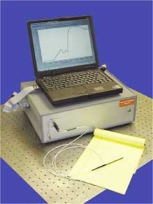
Bigio's current clinical studies are focused on developing low-cost spectroscopic instrumentation and fiberoptic probes for diagnostic applications where there is significant potential volume, such as early detection of cancer of the breast, prostate, lung, colon, and esophagus (see Fig. 3). With breast cancer, for example, he and his colleagues are developing elastic light-scattering spectroscopic probes that can be used both for diagnostics and during surgery, such as detecting whether the cancer has spread to the lymph glands in the armpit and thus improving the prognosis for patients.
"We have been testing our elastic light-scattering spectroscopic technology (which uses a pulsed xenon arc lamp in the near-UV to near-IR) for checking sentinel lymph nodes in the arm pit, which is the first site to become metastatic if cancer is spreading. he said. "The idea is that surgeons could use this to guide the degree of radicalness during surgery."
As with OCT, government funding is helping to push the development and commercialization of spectroscopy for medical diagnostics. Researchers at the George R. Harrison Spectroscopy Laboratory at the Massachusetts Institute of Technology (MIT; Cambridge, MA), for example, are using a $7.2 million NCI/NIH grant to further develop and implement spectroscopic techniques for diagnosing dysplasia in the cervix and mouth. According to Michael Feld, director of the lab, his group has developed a portable instrument that delivers low-level laser energy and white light through an endoscopic fiberoptic probe onto the patient's tissue, analyzing tissue over a 1-mm region. The device has shown promise in clinical trials, identifying invisible precancerous changes in the colon, bladder, esophagus, cervix, and oral cavity.
Even further out
Other optical methods continue to gain ground as well, although they are even further from commercialization than OCT and traditional spectroscopy. These techniques include photon migration, multiphoton excitation, and molecular imaging.
Photon migration involves the use of light in its scattered or diffuse state to acquire images. Although the spatial resolution is generally below that of computed tomography or MRIs, there are numerous advantages, such as low cost, portability, near-zero risk, and the ability to monitor blood oxygenation and blood volume changes in real-time. Clinical applications being explored include breast and brain cancer detection and diagnosis, stroke, brain hemorrhage, brain oxygenation, brain development, muscle oxygenation, and peripheral vascular disease.
Multiphoton excitation was originally studied for use in biomedical applications such as gene inactivation, but researchers now are looking at multiphoton excitation for medical diagnostics. At Beckman Laser Institute, for instance, scientists have been studying this technique for imaging human tissues and their processes, such as coronary arteries and the mechanical properties of these arteries.
"Multiphoton excitation is a very good technique for looking at thick biological tissues," Tromberg said. "One of the exciting developments is second-harmonic imaging using IR short-pulse lasers to monitor the progression of engineered tissues and to image processes like wound healing. We are looking at disease processes in which there is degradation of the extracellular matrix (like cancer) or degenerative diseases where there is scar formation. This is really a very powerful way to look at tissues, both the cells and the extracellular matrix."
The use of fluorescing dyes in conjunction with optical microscopy is a well-developed field. Today, researchers are applying in vitro fluorescent-imaging techniques to small animals in vivo. While this molecular imaging technique is still being used predominantly for drug discovery, the imaging of molecular signatures is expected ultimately to enable earlier detection of disease. In addition, the imaging of specific proteins or pathways will allow clinicians to tailor therapies to individual patients by better drug selection and monitoring noninvasively molecular events that change medical treatment quickly. All of this is expected to result in the creation of a molecular medicine approach to patient care.
"The medical imaging and optical contrast market is going to grow and will top $1 billion in the next five years, potentially reach $10 billion in 10 years," said David Benaron, a teaching professor at Stanford University and founder of five biomedical optics companies—the most recent being Spectros (Portola Valley, CA), which has developed an optical device for identifying ischemia (lack of blood flow to tissue) that should be ready for FDA review this year. "A lot of optics technologies have not been successfully embraced because they were a solution looking for a problem. In our case, we identified the problem first. And when you identify something at the single-molecule level, it means you have done it optically."
REFERENCE
1. T. M. Yelbuz et al., "Optical Coherence Tomography, A New High-Resolution Imaging Tech. to Study Cardiac Development in Chick Embryos," Circulation, 2002;106:2771; circ.ahajournals.org.


