BIOMEDICAL IMAGING: Eye imager quickly shows 80% of retina
A triple-laser retinal imaging device designed to assist secondary eye-care practitioners in diagnosing, monitoring, and treating disorders such as diabetic retinopathy, glaucoma, diabetes, high blood pressure, age-related macular degeneration, and cancer is proving to be more effective than competing imaging systems in detecting disease earlier and potentially saving sight or even lives.
The instrument, developed by Optos (Dunfermline, Scotland), has produced images showing approximately 80% of the retina in a quarter of a second without irritation, according to Tom Daniells, vice president of marketing in North America for Optos. It can also check for leakage, assess tumors, monitor blood flow or oxygen levels, and look for bleeding. The system recently received marketing approval from the U.S. Food and Drug Administration and won the Royal Academy of Engineering MacRobert Award in London in June.
Optos was established in 1992 when the son of the founder, Douglas Anderson, lost the vision in one eye after a retinal detachment was diagnosed too late. Motivated to develop a noninvasive imager that could provide a wider field of view of the retina than the typical 5%, Anderson initially came up with the Panoramic200 (P200) scanning-laser ophthalmoscope, which uses low-power 633 and 532 nm lasers and images the eye through a minimum of a 2 mm aperture. The two lasers are combined into a single beam that is projected onto the patient’s retina and manipulated through a 200° interior angle of the retina.
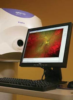
Light reflected from the retina is returned through the scanning system and converted to electrical impulses by highly sensitive photodiodes. These images are digitized and formatted through proprietary software into a high-resolution retinal image called an optomap (see figure). The software enables the practitioner to capture, manipulate, and enhance the image as required, allowing for a detailed evaluation of the retina and producing a permanent clinical record of the examination that can be stored and transmitted to other health care providers.
The P200 contains an ellipsoidal mirror that creates a second focal point behind the patient’s iris to enable imaging from inside the eye, providing pictures of more than 80% of the back of the eye without dilation, according to Daniells. The shape of the mirror provides a better image of the back of the eye because the imaging can take place on the sides of the bowl shape of the eye. Designed for primary practitioners who want to diagnose the overall health of the eye, the P200 has been commercially available since 2000. More than 2000 units are in use, and 7.5 million examinations have been conducted with them in the United States, Germany, the United Kingdom, and Canada.
Building on that technology, the P200MA produces ultrawide, dynamic, high-resolution angiographic images of the retina. The addition of a 488 nm blue laser enables ophthalmologists to perform fluorescein angiography (FA) of the same view, creating a stack of sequential images called optomap fa. The instrument produces a standard retinal exam; an enhanced retinal exam with resolution, eye steering, and zoom capabilities; and FA dynamic ultrawide-field angiography.
Clinical trials included a multicenter pilot study that indicated a statistically significant increase in the mean retinal image area as compared with conventional digital FA imaging with 45% more of the retina being imaged. Enhanced capabilities, including taking pictures quickly with a panoramic view, zooming in to a point of interest, and capturing more of the retina, are likely to help diagnose diseases that manifest themselves in the periphery of the retina and thus identify and treat these diseases faster, according to clinical trial participants.
“The new technology is a more practical and valuable approach than montaged narrow field-of-view images that only provide a composite from different parts of the retina taken at different points during the circulation of fluorescein dye,” Daniells said.