Molecular structure discovery promises rainbow of new probes
"We need to have a lot of colors to study events simultaneously in one cell," says Vladislav Verkhusha of Albert Einstein College of Medicine at Yeshiva University (New York, NY). Today researchers can follow only two or three proteins at a time; an expanded fluorescent protein (FP) palette would advance biological discovery.
Verkhusha is principal investigator of a study describing how Einstein scientists have determined the crystal structures of two key FPs–one blue, one red–used to highlight molecules in living cells. The finding has allowed them to propose a chemical mechanism by which the red color in FPs is formed from blue. With this information, the team now has the first roadmap for rationally designing new and different-colored FPs to illuminate intracellular structures and processes. Colored probes could provide a window into how biological processes in normal cells differ from those in cancer cells.1
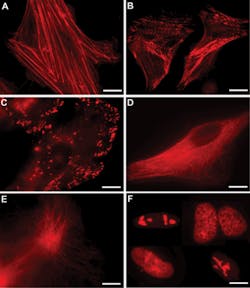
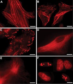
Knowing the structure of the red color in fluorescent proteins promises to allow scientists to develop other colors, and possibly also to improve the brightness of imaging agents. (Images courtesy Kiryl Piatkevich, Yeshiva University)
This is expected to expand the imaging revolution that began with green fluorescent protein (GFP) found in jellyfish, which enabled observation of living cells' internal workings using optical microscopes and won the 2008 Nobel Prize in Chemistry for GFP's discoverers. Many FPs of various colors were later found in other marine organisms, but their molecular nature has remained a mystery, hindering development of new imaging probes. So scientists would fuse genes for known FPs to bacteria, and then expose millions of the microorganisms to radiation, in hopes of producing random genetic mutations that lead to new fluorescent proteins.
- O.M. Subach et al., Chem. & Biol. 17(4), 333–341.
More Brand Name Current Issue Articles
More Brand Name Archives Issue Articles

