BioOptics BreakthRoughs
The latest reseach and technology developments
Photoacoustic tomography images epileptic seizures
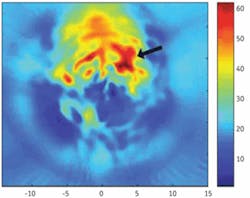
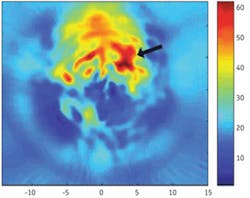
Noninvasive laser-induced photoacoustic tomography (PAT) is an emerging imaging modality that, because of its unique ability to image biological tissues with high optical contrast and ultrasound resolution, has the potential to image the dynamic function of the brain. Huabei Jiang and colleagues at the University of Florida (Gainesville) have demonstrated the use of finite-element-based PAT for imaging of epileptic seizures in an animal model. In vivo photoacoustic images were obtained in rats with focal seizures induced by microinjection of bicuculline into the neocortex. The seizure focus was accurately localized by PAT, as confirmed with the gold-standard electroencephalogram. Compared to the existing neuroimaging modalities, PAT not only has the advantage of high spatial and temporal resolution in a single imaging modality, but also is portable and low in cost, making it possible to bring brain imaging to the bedside. Contact Huabei Jiang at [email protected].
null
Laser-activated nanoparticles detect breast-cancer cells
A novel photoacoustic system combining laser energy and nanotechnology has the potential to detect and quantify circulating breast cancer cells based on in vitro testing of human blood, according to research presented at the American Society for Laser Medicine and Surgery annual conference in April. Spearheaded by John Viator, assistant professor of biological engineering and dermatology at the University of Missouri (Columbia, MO), the breast-cancer cell research models previously published work by Viator and his colleagues in which the laser-activated system (which utilizes a tunable solid-state laser from Optotek) successfully used gold nanoparticles to identify circulating melanoma skin cancer cells. The nanoparticles were used as optical contrast agents or markers to target breast-cancer cells contained within white blood cells. This process is based on the fact that the gold nanoparticles ignore the white blood cells and stick only to breast-cancer cells. Also, white blood cells do not absorb light so they are not illuminated by the 5-ns-long pulsed laser energy used in the study.
However, gold nanoparticles give color to the attached breast-cancer cells, and, when zapped with the laser, the colored breast-cancer cells create an ultrasonic pulse (a high-frequency sound in the megahertz range). An ultrasonic sensor that sends in light and gets sound back in return then listens to the pulses to identify if the nanoparticles (and attached breast-cancer cells) are present. Viator observed that very strong signals were emitted when breast-cancer cells were present, which were detected in clusters of 50 cancer cells. His team plans to begin an animal study in spring 2008 and patient trials in 2009. Contact John Viator at [email protected].
Hyperspectral fluorescence images bacterial pigments during photosynthesis
Scientists at Arizona State Unversity (ASU; Tempe, AZ) are using hyperspectral fluorescence imaging to identify and visualize discrete pigments in live bacteria cells. This approach could help researchers fine tune bacteria for specific purposes, according to Wim Vermaas, a professor in ASU’s School of Life Sciences and lead author of “In vivo hyperspectral confocal fluorescence imaging to determine pigment localization and distribution in cyanobacterial cells” (Proc. Natl. Acad. Sci. USA, 10.1073/pnas.0708090105, Mar. 3, 2008).
Confocal fluorescence microscopy is an excellent method for localizing pigments in cells, as long as there is little spectral overlap between different fluorescing pigments. Hyperspectral fluorescence imaging pushes the boundaries of this technique by separately localizing pigments with similar fluorescence spectra. In experiments conducted by Vermaas and colleagues from ASU’s Center for Bioenergy and Photosynthesis and Sandia National Laboratories, a 488 nm laser was used to excite phycobilins and carotenoids in cyanobacterial cells, yielding fluorescence from phycocyanin, allophycocyanin, allophycocyanin-B/terminal emitter, and chlorophyll a. Resonance-enhanced Raman signals and very weak fluorescence from carotenoids were also observed. The research team found that phycobilin emission was most intense along the periphery of the cell whereas chlorophyll fluorescence was distributed more evenly throughout the cell, suggesting that fluorescing phycobilisomes are more prevalent along the outer thylakoids. Contact Wim Vermaas at [email protected].
FTIR spectroscopy enables predictive models of tumor histopathology
At Pittcon 2008 (March 2–7; New Orleans, LA), Rohit Bhargava from the University of Illinois (Urbana-Champaign) discussed his group’s work with Fourier-transform infrared (FTIR) spectroscopic imaging to automate the analysis and recognition of tissue structure and disease without the need for dyes, molecular probes, or human intervention. According to Bhargava, FTIR imaging is emerging as a preferred technique for high-throughput biomedical sampling applications, combining the molecular selectivity of spectroscopy with the spatial specificity of optical microscopy.
Working closely with researchers from the National Institutes of Health (Bethesda, MD), Bhargava and colleagues are developing a high-fidelity spectrometer that combines a focal plane array detector with a conventional interferometer to potentially increase the speed of data acquisition several-fold. According to Bhargava, they have achieved mathematical noise reduction that enables 50- to 100-fold higher rate of data collection. They also used precise optical modeling to elucidate and resolve various optical effects. Their technique has been used to automate human cancer diagnoses and grading for different tissue types by applying it to routine archival tissue samples, including prostate histopathology, breast tissue histology, and epithelium in lymph nodes. Contact Rohit Bhargava at [email protected].
Phosphate glass exhibits antibacterial properties
Researchers at the University of Kent (England), in collaboration with teams at the Eastman Dental Institute and Warwick University, have produced and characterized a novel phosphate-based glass that exhibits antibacterial properties. Their paper appeared in February in Advanced Functional Materials.
Implant surgery is becoming more common as the average human life expectancy increases. While the ability to repair worn-out or broken body parts is a positive thing, there remains the problem of implant-related infection, including those associated with drug-resistant organisms. These infections can require patients to undergo secondary operations, resulting in additional discomfort, and can even lead to complete device removal. The silver-doped phosphate glass produced in this recent study has a built-in antibacterial mechanism that prevents the growth of bacteria at the tissue-implant interface without interfering with the natural healing process of the body. Silver ions are known to exhibit biocidal characteristics on a large range of bacteria, including MRSA and Clostridium difficle, both of which are major causes of infections in this context. The dissolution rate, and hence silver release, of this glass can be tailored to meet specific requirements, say the researchers. Contact Robert Moss at [email protected].
Metal surface plasmons enhance single-cell fluorescence
The use of fluorophore-metal interactions has the potential to dramatically increase the detectability of single fluorophores for single-molecule detection and fluorescence correlation spectroscopy. Researchers at the University of Maryland’s Center for Fluorescence Spectroscopy (CFS; Baltimore) have been studying the interactions of fluorophores with metallic particles or surfaces and have observed a number of important spectral changes, including increases in intensity and photostability, decreased lifetimes due to increased rates of radiative decay, and increased distances for FRET. They have also shown that fluorophores can create surface plasmons in metals, which in turn create light.
In a paper published March 15 online by the American Chemical Society (ASAP Nano Lett., 10.1021/nl080093z), Joseph Lakowicz, director of the CFS, and colleagues from the School of Medicine demonstrated the role of plasmon-controlled fluorescence in single-molecule counting. Multiple Alexa Fluor 647-conjugated concanavalin A molecules were covalently bound to a single 20 nm silver particle to synthesize metal-plasmon-coupled probes (PCPs). Fluorescence images were recorded using scanning confocal microscopy in both intensity and lifetime. The brightness of the PCPs was 30 times brighter than those of free conA, and the lifetime of the PCPs was shortened dramatically. These results suggest that fluorophore-metal interactions can be used to control the migration of electromagnetic energy across and through metal surfaces, and to control when and where the energy is converted back into light. Contact Joe Lakowicz at [email protected].

