Three-chip HDTV brings new vision to voice disorders

By Gary Pitre
The scientific-imaging, broadcast, factory-automation, and industrial-video markets are discovering the many advantages of using 3CCD (three-chip) technology in diagnostic, video-imaging, and process-control equipment. With recent improvements in high-definition (HDTV) technology and the availability of smaller high-definition video-imaging systems, the applications are growing as quickly as the technology is changing.
Three-chip, HDTV color imagers are now delivering the same quality imagery to the biomedical profession that was once reserved for the broadcast market. The much-improved HD reproduction quality in these devices produces much better color fidelity than was previously available with single-chip color cameras. The truer color, sharper detail, and brighter contrast made possible by these camera systems provides critical information to the diagnostician and researcher and is a tremendous aid to the pathologist and physician in making diagnoses and treatment decisions.
In addition to high-resolution imagery, a key advantage of these next-generation HDTV cameras is the reduced size and weight. They fit comfortably into the available space in a lab, a doctor’s office, or a medical clinic and can be easily transported from one room to another.
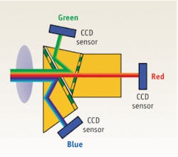
Toshiba Imaging (Irvine, CA), for example, has developed an ultracompact HDTV color camera, the IK-HD1, that can easily be integrated into medical-imaging systems. Toshiba utilizes a proprietary prism block technology; 3CCD imagers contain three separate image sensors and a prism that divides the incoming light rays into their red, green, and blue components (see Fig. 1). Each chip then receives a single color at full resolution, thus providing the best color accuracy available.
In the medical-imaging market, the IK-HD1 HDTV camera is being integrated into a digital video endoscopy/stroboscopy instrument developed by KayPENTAX, A Division of PENTAX Medical Company (Lincoln Park, NJ). The Digital Video Stroboscopy System (the Digital Strobe) is specifically designed for use in the physician’s office. It is an advanced laryngeal imaging system that digitally records stroboscopic and other endoscopic exams to a fully integrated video capture and playback workstation. Originally developed for otolaryngologists, phoniatricians, speech pathologists, and voice scientists, the Digital Strobe provides an analytical imaging system for comprehensive assessment of the vocal cords structure and vibratory characteristics. This same instrument is also being used to record other endoscopic procedures such as transnasal esophagoscopy, which enables the clinician to view the tissue more clearly thereby aiding in more accurate diagnoses.
How stroboscopy works
The basic components of a stroboscopy system are an endoscope, a stethoscopic microphone, a light source, an electronic control unit, a camera, and recording equipment. The microphone is applied to the patient’s neck to extract the fundamental frequency of the patient’s voice while they are phonating during the exam. The extracted frequency is then synchronized with the strobe light to produce a slow-motion rendering of vocal fold physiology. The frequency represents the number of times the vocal cords open and close per second and it is measured in Hz.
The endoscope (coupled to a camera and strobe-light source) is passed transorally or transnasally into the patient’s pharynx, where key anatomical structures are observed during the exam. As the flash of the light is synchronized with the frequency of the patient’s voice, a series of images are recorded allowing the clinician to view any position of the folds during their vibratory cycle to investigate their structure. If the frequency of the strobe is slightly varied with the frequency of the patient’s voice, you will be able to observe the vibratory motion of the vocal folds through continuous glottal cycles.
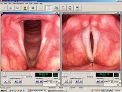
Perceptual judgments of the vibratory patterns of the vocal cords are based on knowledge of the anatomy, the normal gross anatomic appearance of the fold, and the variance of mechanical properties of the fold as a function of frequency and intensity. The Digital Strobe permits the physician to capture clear, streaming video during the examination, without the artifacts. The KayPENTAX system also captures audio (patient audio or the examiners comments) during the procedure. With excellent image quality, the captured data (both stills and MPG and DV (AVI) and MPG video clips) can be played or viewed in other computers, sent electronically to colleagues, or placed in a PowerPoint presentation for future consultation (see Fig. 2).
The crisp, clear stroboscopy images allow the clinician to make an earlier and more thorough diagnosis and provide more appropriate treatment based on the patient’s anatomy and physiology.
ACKNOWLEDGMENT
The author wishes to thank Robert McClurkin of KayPENTAX for his assistance with this article.
GARY PITRE is Eastern Regional Sales Manager at Toshiba Imaging Systems Division (Irvine, CA). Contact [email protected]; www.tais.toshoiba.com.