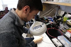
Fiber-optic laser-scanning microscope focuses on brain tissue
A team of neuroscientists and bioengineers at the University of Colorado Anschutz Medical Campus (Aurora, CO) and scientists from The University of Colorado Boulder (CU-Boulder, Boulder, CO) have created a miniature, fiber-optic microscope designed to peer deeply inside a living brain. The researchers published details of the microscope in Optics Letters.
Related Article
"Microscopes today penetrate only about one millimeter into the brain but almost everything we want to see is deeper than that," said professor Diego Restrepo, one of the paper's authors and director of the Center for NeuroScience at CU Anschutz. "You can manipulate this lens while most others are fixed. That means you can see neurons firing inside a living brain."
The laser-scanning microscope, a prototype that will be further refined, uses fiber-optics and a tiny electrowetting lens. Compared to other small, focusing lenses, it’s fast and not sensitive to motion. This allows it to reliably focus on living tissue. At the same time, the lens allows a rapid shifting of focus by applying electricity across two different liquids, which actually changes the curvature of the lens.
About half an inch in diameter, the microscope can be directly mounted onto the head of a mouse. A thin, fiber-optic cord will allow the animal to freely roam while scientists look inside its brain and monitor reactions to certain stimuli. That means parts of the living brain like the amygdala, which had been virtually off-limits to microscopes, will soon be seen in real-time, high-resolution, 3D images.
"Using optical methods to stimulate and record from neurons is the future of neuroscience research," said Baris Ozbay, a doctoral student in bioengineering at CU Anschutz and lead author of the paper. "But most researchers are adapting existing large microscopes to fit mice for head-fixed imaging which limits movement, is difficult to set up and has issues with motion. The solution is to put the microscope on the mouse, rather than putting the mouse on the microscope."
Emily Gibson, assistant professor of bioengineering at CU Anschutz and senior author of the study, said the microscope opens a new world for scientists. "We can now measure a large region and sample more neurons," she said. "For example, we can image up to 100 neurons at the same time, as opposed to perhaps the 10 or so we could do in the past."
All of this has potential human applications as well. "The ability to see beneath the surface of the brain offers new, powerful ways to study brain function," said Restrepo. "It will help us understand brain disease and formulate new treatments."
Other possibilities include screening pharmaceuticals targeted to specific brain disorders, allowing neurosurgeons to image small brain areas like those targeted in the treatment of Parkinson's disease, conducting optical, in situ, biopsies for diagnosis of brain tumors, and examining the connection between neural damage and controlling prosthetic limbs.
Research was made possible by a $1 million grant from the National Science Foundation.
SOURCE: University of Colorado Denver Anschutz Medical Campus; http://www.ucdenver.edu/about/newsroom/spotlight/researchers/Pages/Researchers-create-microscope-allowing-deep-brain-exploration.aspx
About the Author

Gail Overton
Senior Editor (2004-2020)
Gail has more than 30 years of engineering, marketing, product management, and editorial experience in the photonics and optical communications industry. Before joining the staff at Laser Focus World in 2004, she held many product management and product marketing roles in the fiber-optics industry, most notably at Hughes (El Segundo, CA), GTE Labs (Waltham, MA), Corning (Corning, NY), Photon Kinetics (Beaverton, OR), and Newport Corporation (Irvine, CA). During her marketing career, Gail published articles in WDM Solutions and Sensors magazine and traveled internationally to conduct product and sales training. Gail received her BS degree in physics, with an emphasis in optics, from San Diego State University in San Diego, CA in May 1986.