High-resolution OCT probes biological tissue
Noninvasive optical techniques promise to enable in vivo imaging of cells and tissue with a resolution that reveals fine details of morphology. In particular, optical-coherence tomography (OCT) can provide cross-sectional visualization of nontransparent biological tissue if it can attain micron-scale resolution and millimeter-range penetration depth.
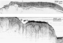
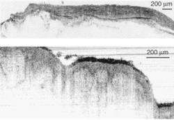
Recently, Wolfgang Drexler and his colleagues at the University of Vienna (Vienna, Austria), along with researchers from MenloSystems (Martinsried, Germany), have tested a broad-spectrum fiber laser emitting at 1375 nm to achieve sub-2-µm-axial-resolution OCT in nontransparent biological tissues.1 This resolution represents a 25% increase in axial OCT resolution. The tissues were ex vivo samples from a human artery and skin, which were imaged through thin layers of water to prevent dehydration and to optimize the index matching (see Fig. 1).
Optical-coherence tomography provides cross-sectional images of biological structures analogous to ultrasound. Whereas ultrasound reads the backscatter of sound waves and produces a relatively low-resolution image, OCT relies on variation in the coherent backscatter of light to generate a relatively high-resolution image. Any physical property that changes the amplitude, phase, or polarization of the backscattered light could result in information of diagnostic value. A simplified OCT system consists of a low-coherence interferometer that is fed by a light source. The source beam is split into two paths: the reference and the sample. The reflected beams return to the interferometer and generate a cross-correlation signal that is directed toward a detector and signal processing equipment.
Light sources
Numerous light sources have been developed for OCT. Narrowband superluminescent diodes have provided axial resolution of 10 to 20 µm. Submicron axial resolution has been achieved by broadband photonic-crystal-fiber and thermal sources, although the image-penetration depth has been limited to a few hundred microns. And 1-µm axial resolution has been achieved with better depth resolution in the 800-nm region by using Ti:sapphire femtosecond lasers.
In the case of the Austrian researchers, the broadband light source covered wavelengths from 1200 to 1800 nm. It is based on a pulsed, modelocked erbium-doped fiber laser and dispersion-shifted fiber that is highly nonlinear, acting as the medium to broaden the output spectrum of the laser. The fiber laser emits pulses shorter than 200 fs with an average power of a few milliwatts. To achieve high nonlinearity, the pulses are amplified to an average power of almost 100 mW before being guided through the fiber. By operating the system in the 1300-nm region, the OCT system obtains both high-resolution and a penetration depth of several millimeters.
To avoid spectral losses, the researchers chose to interface the filtered output of the fiber laser source to a free-space OCT system to demonstrate the full potential of its broad emission bandwidth. All the optical components of the OCT system were selected to support the propagation of broad-bandwidth light in the range of 1100 to 1600 nm. The filtered fiber-laser spectrum was moderately modulated and centered at 1375 nm, with a full width at half maximum of 470 nm and a power output of 4 mW. However, only 0.5 mW of the 4 mW reached the sample surface because of power loss at the polarizing beamsplitter caused by the randomly polarized output of the fiber laser.
Individual layers and membranes are clearly visible in the artery and skin samples. The researchers believe this shows that an OCT system based on such a stable, low-cost, compact fiber laser has great potential for in vivo OCT clinical studies. To reach this point, laser performance must be improved by smoothing the emission spectrum and increasing the system output power two- to fivefold. A fiber-based OCT system capable of supporting the broad spectral output of the fiber laser will also have to be developed.
REFERENCE
- K. Bizheva et al., Optics Lett. 28(9) (May 1, 2003).