A microscope designed around recoverable blur?
For decades, scanning microscopy has treated motion blur as a defect to be prevented. The familiar stop-and-stare workflow reflects this assumption: Move the specimen, halt the stage, wait for it to settle, expose the camera, and repeat. While it’s a reliable way to protect image quality, the repeated acceleration, settling, and exposure steps also make the mechanics carry much of the system burden.
Computational imaging invites a more adventurous question: If the blur produced by continuous motion is structured and reproducible, can an algorithm recover the information needed for a defined task? It doesn’t make blur desirable in itself, but rather treats it as a precise engineering variable that can be considered alongside stage speed, exposure time, optical resolution, signal-to-noise ratio, training data, and the cost and complexity of the hardware.
Motion blur as a systems-engineering design choice
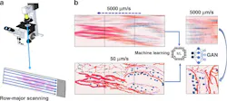
In earlier work with the late Professor Gabriel Popescu, we tested this idea in GANscan, a continuous-scanning method.1 Rather than stopping the stage for each field of view, the microscope recorded video while the specimen moved. A conditional generative adversarial network (GAN) was trained on registered pairs of motion-blurred and sharp images. Our published experiments used a conventional microscope without specialized scanning hardware and demonstrated restoration at stage speeds up to 5,000 µm per second. In this experimental setting, acquisition throughput was reported at up to 30x the stop-and-stare comparison, while inference on 256-by-256-pixel frames took less than 20 ms on a consumer graphics processing unit (GPU).
Our results established a proof of principle, not a universal permission slip for neural restoration. A network can only be judged against the data, specimen types, optics, motion, and failure modes represented in its validation. The GANscan study compared restored images with slowly scanned and fully stopped controls, evaluated spatial-frequency recovery, and tested tissue from patients outside the training set. It also examined defocus and showed that reconstruction quality declined as the focal offset increased. The limitation is part of the design result: Computation can relax some constraints, but it does not erase physics.
The physics makes the tradeoff concrete. During an exposure, a translating specimen is integrated across the distance traveled by the stage, so the smear grows with velocity and exposure time. At sufficiently high motion, spatial frequencies disappear rather than merely become less contrasty. Classical deconvolution can narrow broadened features, but it can’t recreate information not recorded. A learned model can predict plausible high-frequency structure from examples, which is useful only when that prediction is tested against appropriate controls. For system designers, this means the blur distribution used in training must follow from the actual stage motion, optics, camera integration time, and specimen preparation—not from a generic image-processing recipe.
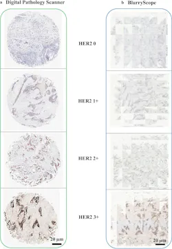
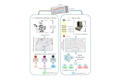
The next question was whether this acquisition philosophy can be embodied inside a compact, task-specific research platform. Yes—our team at the Bio- and Nano-Photonics Laboratory at UCLA developed BlurryScope in 2025,2 and the prototype used continuous brightfield scanning, automated stitching and cropping, and neural networks for HER2-score classification from motion-blurred images of immunohistochemically stained breast tissue microarrays. The instrument measured 35-cm tall, weighed 2.26 kg, and used low-volume components at a total cost of $450 to $650.
Our HER2 study was intentionally demanding because stain intensity and membrane continuity both matter. The blinded test set comprised 284 unique patient cores, each scanned three times. We reported 79.3% accuracy for four-class scoring and 89.7% for a grouped two-class task under the specified analysis. These figures should remain attached to the exact study design. BlurryScope was presented as a proof-of-concept research system with explicit tradeoffs in resolution, signal-to-noise ratio, and the smallest detectable features—not as a replacement for general-purpose pathology scanners or a standalone diagnostic device.
Codesign acquisition and reconstruction
For optical engineers, the broader lesson is methodological. Acquisition and reconstruction should be codesigned rather than optimized in isolation. Increasing stage speed changes the blur kernel and the spatial information available to the model. Shortening exposure can reduce blur, but may sacrifice photons. A more compact mechanical architecture may improve accessibility, while narrowing the range of samples or tasks it can support. The appropriate design point depends on the information the user actually needs and how reliably the system can recognize when it is outside its validated domain.
A practical architecture should expose, rather than conceal, the variables that connect the physical scan to the model. Stage velocity and direction, exposure, focus, objective, illumination, and preprocessing should be recorded with the image data. Quality checks can then ask whether a new frame resembles the validated acquisition envelope before reconstruction or analysis proceeds. This kind of traceability is less visually exciting than a before-and-after image, but it turns a demonstration into an engineering system that can be characterized, reproduced, and improved.
It also changes how performance should be communicated. An attractive reconstruction isn’t enough. Developers should preserve sharp controls, separate training and patient-level test sets, report indeterminate cases, probe focus and motion sensitivity, and evaluate repeatability across independent scans. Task-specific accuracy and image-restoration fidelity answer different questions and shouldn’t be blended into a single claim. Where the application is biomedical, the distinction between a peer-reviewed research finding and a commercial product statement is especially important.
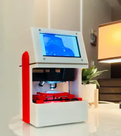
To translate this research work into a product, I launched FanousPhotonics (SCANIMUS is our research-use-only product). While UCLA and the Bio- and Nano-Photonics Laboratory are the provenance of the research, it isn’t a product endorsement.
One way to think about deliberate blur is not as a software trick that rescues weak hardware, but as a careful systems-engineering choice. When motion is controlled, training data are representative, controls are rigorous, and the task is carefully bounded, computation can redistribute complexity across optics, mechanics, electronics, and algorithms. Sometimes the right answer will still be to stop the stage. In other cases, accepting a measured amount of imperfection at acquisition may be exactly what makes a faster or more accessible architecture possible.
DISCLOSURE
The 2025 BlurryScope publication discloses pending patent applications related to the research.2 UCLA, the Bio- and Nano-Photonics Laboratory, and the cited publications are not represented as endorsing FanousPhotonics or SCANIMUS.1,2
REFERENCES
1. M. J. Fanous et al., Light Sci. Appl. (2022); https://doi.org/10.1038/s41377-022-00952-z.
2. M. J. Fanous et al., Dig. Med., 8, 506 (2025); https://doi.org/10.1038/s41746-025-01882-x.
About the Author
Michael Fanous
Michael Fanous, Ph.D., is the founder & CEO of FanousPhotonics, which is developing SCANIMUS as a research-use-only product. He is the lead author of the peer-reviewed GANscan and BlurryScope papers.