High-index microsphere layer on coverslip performs super-resolution microscopy
Researchers from the Department of Radiation Oncology at the University of Pennsylvania (Philadelphia, PA) are using microscope coverslips composed of a monolayer of microspheres with high refractive index positioned in a transparent elastomer layer for simple, generic super-resolution imaging that improves the resolution of optical imaging by a factor of 2 to 3.
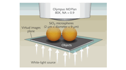
The research team fabricated several different microscope coverslips by spin coating barium titanate glass (BTG) microspheres (refractive index 1.9-2.1) in a polydimethylsilaxane (PDMS) layer whose thickness can be controlled by the spin speed and duration. When the slide is placed over the specimen, a magnified virtual image is formed by the microsphere and captured by the objective lens. Improving the spatial resolution by microsphere-assisted imaging acts by enhancing the effective numerical aperture (NA) of the system and maximizes the extraordinary focusing properties of microspheres via the so-called photonic nanojet effect. It should be noted that the focusing properties of the microsphere-embedded films can find applications in other areas such as nanoscale patterning, spectroscopy, and photovoltaics.
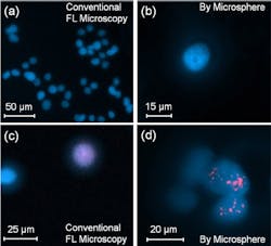
The researchers also investigated the feasibility of using their microsphere-assisted technique to image biological structures, such as cells and tissue sections. A limiting factor in predicting patients' response to radiation therapy is the sensitivity of their tumor cells to ionizing radiation. This sensitivity can be assessed by observing the repair of double strand breaks (DSB) in DNA, which can be detected via phosphorylated histone, a highly specific and sensitive molecular marker. Cells are exposed to radiation, and the number of histone foci induced in the cell nucleus is measured as a function of time after radiation exposure. The disappearance of foci correlates with the repair of the DSBs. Standard microscopy does not provide sufficient resolution to characterize the quality of the foci. Microsphere-assisted imaging can be used in histone assays to obtain enhanced images that overcome this limitation.
The team also investigated using microsphere-assisted imaging for tissue sections by imaging mouse kidney tissue sections to analyze immunostaining of glomeruli, which serve as the kidney's filtration units. Various glomerular diseases primarily target the glomerular filtration that controls the kidney's ability to filter blood. This requires higher resolution than conventional microscopy provides, which is usually achieved through electron microscopy. The team used FL microscopy and BTG microspheres immersed in a gold antifade mounting solution to image the distribution pattern of antibody-immunostained podocyte protein markers known as ZO-1 and Myo1c. It is known that these proteins are critical for maintaining podocyte structure and function, and their loss may severely affect kidney function. Thus, finding these proteins at the podocyte cell membrane indicates a healthy podocyte. Comparing conventional and microsphere images shows significant improvement by imaging through the microsphere.
The researchers are currently working on further advancing this technology, including optimizing the imaging capabilities and developing a large-scale fabrication technique. We are also expanding the application of microsphere-assisted imaging in cancer and medical sciences.
SOURCE: SPIE Newsroom; http://spie.org/x113361.xml?highlight=x2416
About the Author

Gail Overton
Senior Editor (2004-2020)
Gail has more than 30 years of engineering, marketing, product management, and editorial experience in the photonics and optical communications industry. Before joining the staff at Laser Focus World in 2004, she held many product management and product marketing roles in the fiber-optics industry, most notably at Hughes (El Segundo, CA), GTE Labs (Waltham, MA), Corning (Corning, NY), Photon Kinetics (Beaverton, OR), and Newport Corporation (Irvine, CA). During her marketing career, Gail published articles in WDM Solutions and Sensors magazine and traveled internationally to conduct product and sales training. Gail received her BS degree in physics, with an emphasis in optics, from San Diego State University in San Diego, CA in May 1986.