People often forget that stress is not just a human condition. When subjected to some change in the environment—be it smog, pesticides, global warming, or air or water pollution—even plants will react physiologically or biochemically. Actions include either adapting to that change or reacting in a way designed to protect their organic life systems. The good news is that these adjustments are often followed by changes in absorbance of a plant's leaves, which means that fluorescence parameters can be measured to examine just how stressed the plants really are.
For some time now, spectroscopy has been a principle means used to derive information about how plants react to stress. There is room for improvement, though, namely because of the nature of the reactions. According to researchers at the US Department of Agriculture's Beltsville Agricultural Research Center (Beltsville, MD), most plant leaves illuminated with ultraviolet (UV) radiation will exhibit a broad fluorescence emission with a maximum at 450 nm and a shoulder peak at 530 nm. There also are narrower chlorophyll-related emission bands at 680 and 740 nm. Changes in emission related to changes in stress levels are often presented as ratios of two-band combinations of blue, green, red, and far-red regions of the spectrum. To top that, the mechanisms and plant constituents governing these changes can be quite complex, with the effect on fluorescence in the blue-green band especially obvious.
Multispectral imaging
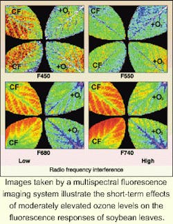
While several recent research projects have used laser-induced fluorescence techniques to demonstrate the feasibility of fluorescence measurements from airborne platforms, these systems often use lasers with some pulse-to-pulse peak power variations. More accurate measurements may some day be possible with multispectral imaging systems, especially those that rely on continuous-wave excitation sources. In line with this, the Beltsville Center scientists, working with colleagues at the University of Maryland (College Park, MD), and NASA's Goddard Space Flight Center (Greenbelt, MD), have developed a steady-state imaging system that can capture fluorescence images of targets such as plant leaves at four spectral bands in the blue, green, red, and far-red regions of the spectrum, which are centered around 450, 550, 680, and 740 nm, respectively.1 While the current configuration is still lab-based, the device can provide qualitative and quantitative characterization of spectral fluorescence changes in a plant, including multiple-band ratios resulting from biochemical and physiological changes, as well as spatial variation information across the plant's surface that is not readily available with integrated-point-source measurements such as provided with pulsed-laser devices.
The imaging system used in leaf analysis incorporates a UV excitation source, a sample holder, interference filters, a CCD digital camera, 12-bit-resolution analog-to-digital converter, and converter interface for instrument control and data collection. The excitation source consists of four 12-W long-wave UV-A fluorescent lamps located 0.2 m above the sample surface at an angle, two per side, that are directed toward a central target area. The UV excitation intensity at the target is 0.33 mW cm-2 with an emission maximum at 360 nm. The radiation from the lamps is filtered to eliminate wavelengths greater than 400 nm and prevent pseudo-fluorescence from the light source.
The front-illuminated CCD camera has spatial resolution of 192 x 165 pixels and provides average quantum efficiency greater than 20% within a band from 400 to 800 nm, with the maximum efficiency found in the 650 to 750-nm region. The Nikon lens mounted to the camera head, with f-1/3.5 and a 20-mm focal length, couples to an automated filter wheel that can hold up to five circular filters, although four were typical for these experiments.
The scientists tested the imaging apparatus on soybean leaves, which exhibit significantly different florescence emission characteristics between their adaxial and abaxial surfaces (see figure). Experiments imaged leaves treated with herbicides, as well as leaves grown in an elevated tropospheric ozone (O3) environment. According to the researchers, the results indicate that instruments measuring fluorescence from an entire leaf surface can give a more accurate assessment of plant health than techniques that are limited to separate integrated-point measurements across the area of the leaf.
REFERENCE
- M. S. Kim et al., Appl. Opt. 40, 157 (Jan. 1, 2001)
About the Author
Paula Noaker Powell
Senior Editor, Laser Focus World
Paula Noaker Powell was a senior editor for Laser Focus World.
