Mobile holography system records 3-D portraits for planning surgery
Holography systems have traditionally been bulky, requiring heavy optical tables. But now that pulsed compact narrowband laser sources are available, such systems can be made small enough to be transportable to the scene that is to be recorded. Furthermore, if the pulse energy is sufficiently high, the exposure can be restricted to a single shot. The hologram carries true 3-D information on the shape of the object, as far as radiation has been backscattered from its surface to the holographic film where it is superposed with the reference beam.
A group led by Peter Hering, University of Düsseldorf (Düsseldorf, Germany) and research center Caesar (Bonn, Germany), takes advantage of those properties for in situ 3-D imaging of faces of patients who are, for example, to be subjected to maxillo-facial surgery.
The one-shot holography does not suffer from drawbacks such as patient motion during exposure. The goal is to gain digital 3-D data on the shape and the skin texture of the face so that the portrait can be visualized in varying directions on the computer screen, and modeled so that facial surgery objectives and results can be directly compared and documented.
Eye safe
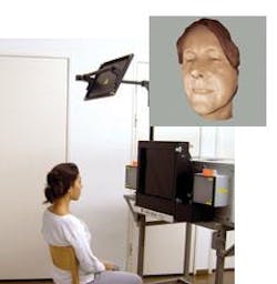
The holographic setup was unveiled at Laser 2005 in Munich (see figure). The laser is a 1.4-J Nd:YAG emitting 1.4-J pulses with 20‑ns duration at a wavelength of 532 nm; its spectral bandwidth results in a coherence length of 6 m. The large coherence length can allow the inclusion of side-mirror views of an object into the same hologram, and the ability to take holograms of much larger objects—for example fine art or even excavations at original archeological sites. The irradiation of patients is done at a low enough level to be eye-safe.
The holograms are recorded on fine-grain silver-halide film that allows up to 4000-lines/mm resolution so that fine details such as pores and hairs can be resolved. Using reconstruction light at 532 nm, the holograms project real images (that is, images that are in focus in front of the hologram); moving a semitransparent screen through the real image allows the 3-D image to be scanned tomographically, slice-by-slice, and digitally photographed. Each picture contains pixels of focused radiation (those that correspond to points on the focused real image) and blurred regions, which are distinguished by a computer algorithm based on gray-value variance.1 The multitude of sharp pixels makes up the 3-D data manifold of the object surface. Until now, direct digital recording of the hologram and numerical reconstruction has failed when such a high resolution was required.
In a screen shot of a head portrayed in this way, fine surface texture can be seen that gives rise to the impression of reality (see figure). The image is easily rotated, allowing it to be seen from the side to as far as the hologram recorded it. Once digital portrait information is available, any face variation can be modeled, for example to plan a surgery. More important, the actual face surface can be correlated to an x-ray tomogram of the underlying bone structure so that any surgery affecting the bones—for example, at the jaw or skull—can be linked to its effect onto the face. Furthermore, if information on crania only is available, faces can be digitally modeled.
REFERENCE
1. A. Thelen et al., J. Opt. Soc. Am. A 22(6) 1176 (2005).
About the Author
Uwe Brinkmann
Contributing Editor, Germany
Uwe Brinkmann was Contributing Editor, Germany, for Laser Focus World.
