Cooled cameras help discover drugs

Charles Slaughter
Large, blue-enhanced two-dimensional CCDs cooled to cryogenic temperatures are central to high-throughput screening of chemical compounds for potential therapeutic drugs.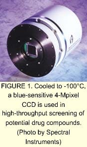
Of the seemingly endless variety of organic compounds, only a select few have potential use as therapeutic drugs. The process of finding these few compounds is termed drug discovery. As in any burgeoning technical field, drug discovery has produced its own set of terms: genomics, proteomics, leads, and screening are just a few. Drug discovery has evolved into a numbers game, with the increased use of combinatorial chemistry as the modus operandi for mix-and-match processes that parade libraries of chemical compounds past ranks of genetic proteinsany of which might reveal compounds that could lead to therapeutic drugs. As the biometrics industry has sought to shorten the time to find and develop new drugs, the technique of high-throughput screening (HTS) has moved to the forefront. Essential to this technique are high-quality imaging systems.
A plateful of wells
While a variety of media can be used to develope leads to new drugs, the micro-titer plate is the most popular. A micro-titer plate is a formed plastic tray 3.5 x 5 in. in size and 3/8 in. thick. Wells are formed as a regular grid of depressions into the top surface of the tray. These plates are available in 96-, 384-, and 1536-well configurations, and in black, white, or transparent material. The 96-well plate is formed as an array of 8 x 12 pockets that are 0.25 in. in diameter. The 96-well array is the easiest to use, as its large wells permit use of cheaper and more user-friendly filling and handling equipmentalthough the wells do use more of the expensive compound materials. The larger arrays (384 or 1536) have smaller wells, which require a higher reading resolution and readers that are more sensitive.
Central to raising the speed of HTS are design improvements that enable the performance of tasks once done serially to be done in parallel. The activity of filling and processing a plate lends itself to automation; a wide variety of plate-handling equipment has been developed both to combine compounds and to evaluate the results. In a serial process, a single photomultiplier reads each cell of a plate placed on a translation stage that sequentially positions each well beneath the photomultiplier. Although this process was at one time sufficiently fast, it has since evolved into the imaging of entire plates using a two-dimensional (2-D) cooled charge-coupled-device (CCD) imaging camera system.
By reading the plates as a whole, the screening process speed has been increased by at least a factor of 100. The combination of highly sensitive 2-D imaging cameras with specialized optics and automated plate stackers has allowed instrument suppliers to produce equipment that processes even high-density plates in a matter of minutesor as many as 100,000 measurements per day.
Seeing with high sensitivity
High-throughput screening involves inspection of the results of every compound testedpeering down into each well simultaneously to "see" what happened. Seeing in this case means measuring the light emitted from each well. Light is emitted by material in the wells through one of three different mechanisms: laser-induced fluorescence, radioactive scintillation, or chemiluminescence. Of these, radioactive scintillation and chemiluminescence are typically low-level emissions predominantly at wavelengths shorter than 460 nm. The laser used to induce fluorescence lights up the entire plate, which then fluoresces wherever there are no wells. Thus, the fluorescence image is typically composed of weak signals within the wells against bright spots from the plate itself.
The requirements for a camera that enables HTS on weakly emitting samples are stringent. Every photon is precious, so the sensitivity must be high. This need translates into CCD imaging systems that are nearly photon counters. Such an action is achieved by using a camera that captures all incoming photons and does not dilute them with thermal photons from within the CCD, nor confuse them by means of noise added by the readout process.
A well-reading detector for all the above luminance mechanisms requires high sensitivity with a quantum efficiency above 80%, low system noise that is less than four electrons, and a dynamic range of 30,000 to 1, or better. A camera developed by Spectral Instruments that fits these requirements contains a blue-sensitive CCD that has at least 1024 samples across the plate. Designed to run at -100°C to eliminate background noise, this high-dynamic-range camera is used in a variety of HTS instruments. With such a camera, plate reading equipment can support a higher well density such as that found on the 1536-well plate.
Systems Integration Drug Discovery Company (SIDDCO; Tucson, AZ) has developed its own approach to gene-based compound screening. The SIDDCO process incorporates a proprietary micro-plate based assay. It uses a standard 96-well plate that includes a 16-element DNA chip at the bottom of each well.
Each element in the chip measures one gene. Thus, each sample is tested for 16 activities at the same time. This Multi-Array Plate Screening (MAPS) protocol can be used to detect DNA, RNA, or both. Unlike larger DNA arrays designed to monitor thousands of genes in relatively large and sparse samples, MAPS is optimized to quantify selected gene sets in multiple small samples with exceedingly high sensitivity and reproducibility. The MAPS transcription assay for quantitatively profiling the expression of 16 different mRNA species can be developed and validated by one person in less than three weeks. Furthermore, establishing a MAPS transcription assay only requires partial sequence information of the target mRNA. Substituting one mRNA target for another is easily achieved since MAPS plates are prepared in small batches.
Demanding requirements
The MAPS process utilizes standard 96-well plates that are supported by a wide variety of plate handling equipment. The plate handling equipment is low-cost, the plates themselves are cheap and easy to obtain and use. However, even though the MAPS process uses the dominant 96-well plate format, its imaging requirements are as demanding as those for 1536-well plate imagers.
The SIDDCO system contains a CCD imager that simultaneously quantifies all the data from the expression profile. The imager used is equipped with a blue-sensitive CCD having a 2048 x 2048 array of 13.5-µm pixels (see Fig. 1). This camera is cooled to cryogenic temperatures using a closed-cycle cryogenic refrigerator to eliminate thermal background. The 16-bit digitizer is operated in a dual-slope integration mode and has a read noise of less than 3.0 electrons. The signal capacity of each pixel is 120,000 electrons, which provides a dynamic range of greater than 34,000 to 1, or 90 dB in a single frame. The system has a sample resolution of 62 µm at the plate. This resolution translates into greater than 100 samples across each 0.25-in. well and provides six samples across each of the 16 sites on the biochip.
In an assay-profiled steroid-induced change in the expression of 13 genes in a cell line, SIDDCO's process reliably detected as few as 30,000 copies of target mRNA in a sample without requiring amplification (see Fig. 2). Detection levels this low are possible due to the high sensitivity of the camera and its wide dynamic range. At 150,000 target molecules per sample, measurements had a standard deviation of less than 10% between replicate samples.
The location of signals within each cell array identifies which target is measured. The light intensity of each spot indicates how much target is present. Imaging and analysis throughput is approximately two minutes per plate. Once the plates are screened, the chemical structures of the active or positive test provide scientists with clues about the characteristics that must be built into the final drug.
CHARLES SLAUGHTER is an applications scientist at Spectral Instruments Inc., 1802 West Grant Road, Suite 110, Tucson, AZ 85745; e-mail: [email protected].