Ultrafast Lasers/Bioimaging: Ultrafast laser pulses enable promising new x-ray imaging
A method based on ultrafast laser-generated x-rays has proven able to generate 3D images of ultrafine structures in the body of a living organism for the first time. The approach could make future medical applications more cost-effective and space-efficient.1
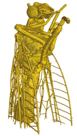
The development includes German researchers from Ludwig-Maximilians University, the Max Planck Institute of Quantum Optics, and Technical University of Munich (TUM). With laser light, researchers from the Laboratory of Attosecond Physics at Max Planck Institute and TUM built a miniature x-ray source able to capture ultrafine details. When physicists Prof. Stefan Karsch and Prof. Franz Pfeiffer illuminated a tiny fly with x-rays, they were able to image the finest hairs on its wings. Coupling the laser-generated x-ray technique with phase-contrast x-ray tomography enabled a 3D view.
The x-rays required for this approach are generated by electrons accelerated to nearly the speed of light over a distance of approximately 1 cm—by laser pulses of ~25 fs with a power of approximately 80 TW. Until now, such radiation could be produced only in enormous, expensive ring accelerators.
Because the technology can distinguish differences in tissue density, it is particularly interesting for medical applications: Cancer tissue, for example, is less dense than healthy tissue. If the researchers are able to shorten the wavelength of the x-rays to penetrate thicker tissue, the method could enable very early cancer detection by imaging tumors less than 1 mm in diameter.
1. J. Wenz et al., NatureCommun., 6, 7568 (2015); doi:10.1038/ncomms8568.
About the Author

Barbara Gefvert
Editor-in-Chief, BioOptics World (2008-2020)
Barbara G. Gefvert has been a science and technology editor and writer since 1987, and served as editor in chief on multiple publications, including Sensors magazine for nearly a decade.