Multimode holographic waveguides tackle in vivo biological imaging

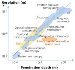
The ability to image biological structures and tissues deep within the human body is met with varying levels of success through such methods as optical coherence tomography (OCT), multiphoton microscopy, and endoscopy. Challenged by the tradeoff of imaging resolution vs. depth, a researcher at the University of Dundee (Dundee, Scotland), with support from the Scottish Universities Physics Alliance (Glasgow, Scotland), has built on earlier work to improve understanding of holographic light transport processes through optical fibers, enabling in vivo imaging through flexible multimode fibers—a feat not previously possible.
Because today's flexible endoscopes with 1 mm diameter or larger fiber bundles are too invasive for sensitive tissues, hair-thin multimode optical fibers were selected to transmit image information along the fiber via several concurring optical modes propagating at different phase velocities. Although the output optical fields are randomly appearing superpositions of these modes with highly complex phase relations, digital holography can characterize these scrambled images empirically, revealing the transmission matrix of the optical system and the details of the image. The most common approach is based on phase-shifting of the input modes such that they interfere constructively at a selected point behind the fiber output facet, thus creating a diffraction-limited focal point. A matrix of such focal points is then used to raster-scan the object to form its image. Currently available components can be used to achieve sub-micron resolution with typically tens of kilopixels at a few frames per second, but the performance is likely to grow steeply in the near future. Reference: M. Plöschner et al., Nat. Photonics, 9, 8, 529-535 (2015).
About the Author

Gail Overton
Senior Editor (2004-2020)
Gail has more than 30 years of engineering, marketing, product management, and editorial experience in the photonics and optical communications industry. Before joining the staff at Laser Focus World in 2004, she held many product management and product marketing roles in the fiber-optics industry, most notably at Hughes (El Segundo, CA), GTE Labs (Waltham, MA), Corning (Corning, NY), Photon Kinetics (Beaverton, OR), and Newport Corporation (Irvine, CA). During her marketing career, Gail published articles in WDM Solutions and Sensors magazine and traveled internationally to conduct product and sales training. Gail received her BS degree in physics, with an emphasis in optics, from San Diego State University in San Diego, CA in May 1986.