Imaging PMTs detect in all dimensions
Imaging PMTs detect in all dimensions
Combination of single-photon counting, x-y imaging, and picosecond time resolution makes imaging photomultiplier tubes useful in advanced applications.
Michael Mellon
Converging technology trends are placing new demands on high-performance scientific detector systems used in ultra-low-light imaging and spectroscopy. Understanding the characteristics and advantages of these detector systems is key to optimizing measurement solutions in the most demanding and sophisticated applications.
For several reasons, many specialized scientific instruments are generating weaker optical signals. Constantly shrinking electronic-device and other analytical sample geometries accompanying increased circuit complexity and speed requirements are leading to smaller elemental optical emitting volumes. Certain types of samples, in particular in biology and biomaterials research, require lower excitation fluxes to avoid sample damage, which also also leads to lower detector signals. Useful analytical techniques of significantly increasing interest, such as luminescence and Raman spectroscopy, are often inefficient producers of photons and demand the highest-sensitivity and lowest-noise detectors.
Because of these weaker signals of interest, relatively longer integration times are often required to achieve suitable signal-to-noise (S/N) ratios to enable the signal of interest to be "seen." This constraint requires a detector with very low detector-generated noise (in which the detector noise contributions are small compared to the photon statistics, even at ultralow photon flux rates). This, in turn, dictates use of a quantum-limited performance detector with the ability to satisfactorily integrate for extended periods without encountering either detector count saturation or interference from extraneous high-energy events such as cosmic rays or other radiation events (for example, from trace radioactivity in detector packaging). Such characteristics are a significant problem in applications of on-chi¥integrating-type detectors such as charge-coupled devices (CCDs) and related devices, and they effectively limit the maximum useful integration time of these devices.
The need for spatially resolved information from nonhomogeneous media and materials in both spectroscopy and imaging is steadily growing. This information must be obtained without resorting to tedious and time-consuming point-by-point, mechanically scanned spatial measurements made with a conventional single-channel detector.
There is also a growing need to make more measurements and simultaneously acquire more data more quickly. In spectroscopic applications, this requirement has led over the past decade to the increased use of one-dimensional (1-D) parallel multichannel detectors (so-called array detectors) that offer enormous gains in data throughput per unit time by processing photons at multiple wavelengths without mechanical scanning. This feature is often referred to as the multiplex advantage of multichannel detection, which is important not only in reducing the time cost of measurements, but also because some samples significantly change characteristics over the longer times required to make measurements in more traditional ways. Use of full 2-D detectors for simultaneous parallel acquisition of data in all pixels of an image has brought time-saving advantages to full x-y imaging applications relative to point-by-point measurements.
Finally, there is growing interest in the observation and characterization of dynamic, transient processes, driven by a wide and diverse range of applications. These include understanding charge transport phenomena in semiconductor materials, spatially localizing defects by optical photon emission microscopy and diagnostically identifying individual device switching times in complex, high-density microcircuits, speckle imaging in astronomy to eliminate the effects of atmospheric turbulence, and research in materials and biomaterials, particularly proteins, using time-resolved spectral fluorescence phenomena. Each of these applications requires combining ultralow-light parallel multipixel and multichannel imaging detection capability with subnanosecond (100 ps or better) single-photon time resolution.
Essentially, what is needed for these applications is a detector that combines the single-photon sensitivity, extremely low noise, and precise time-resolution capability of a photon-counting, microchannel-plate (MCP) photomultiplier tube (PMT) with the full imaging capability of a 2-D detector. Until the introduction of the Quantar Technology 2600 Series Mepsicron multipixel photon imager system, a position-sensitive, single-photon-counting, time-resolving PMT, such a detector has not been routinely available.
Operation of Mepsicron imager
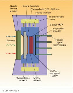
The front section of the vacuum-based Mepsicron imager--a multiple-element position-sensitive imager with time--is similar to a proximity-focused MCP-based PMT operated in a single-photon-counting mode (see Fig. 1). In operation, single incoming optical photons strike the photocathode, generating photoelectrons. In turn, these photoelectrons impact the MC¥electron multiplier surface, physically located directly under the photocathode, under the influence of a strong electric field. At this position, the photon image is replicated, photon by photon, as an electron image on the MC¥surface.
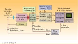
The near-noiseless charge amplification of the MCPs cause the single photoelectron to quickly build into a charge cloud consisting of approximately 107 electrons. Here the resemblance to an image intensifier ends. The gain is considerably higher due to the use of multiple MCPs, and no phosphor screen is used, which avoids problems like optical conversion efficiency, blooming, and phosphor decay time lag. The amplified charge cloud is intercepted by a proprietary charge-division x-y position encoder located behind the MC¥stack. After electronic processing, the x-y spatial position of each incoming single photon is recovered with a position resolution exceeding 50 µm FWHM. The x and y coordinates of each photon are then histogrammed in an external dual-ported digital RAM memory and transferred to a PC-based multiparameter data system for display and analysis (see Fig. 2).
Reconstructed digital 1-D spectra or full 2-D multiple spectra or images are developed, composed entirely of an ensemble of single-photon events. Only events exceeding a preset threshold are counted, thus eliminating much of the background noise of other detection approaches. Because the low intrinsic background count rate from the MCPs and the thermoelectrically cooled photocathode of the device are distributed over the digitized pixels in a 2-D image (typically 800,000 to 1,000,000), the dark count per pixel is remarkably small, on the order of 10-5 to 10-6 counts per second per pixel, an almost negligible level in even the lowest photon count-rate applications and orders of magnitude lower than imaging detectors such as CCDs, even when operated at liquid-nitrogen temperatures.
There is no count-related readout noise such as found in other detectors, resulting in extremely high detective signal-to-noise (S/N) ratios at ultralow photon flux rates, compared to other approaches. In many applications, the average S/N ratio in a single pixel relative to detector-noise background will typically exceed 10,000:1. The Mepsicron offers true single-photon count sensitivity (the user can actually "see" single photons detected in an individual pixel). Such sensitivity is not usually possible with CCD-type detectors due to greater dark count and readout noise, which mask single photons in the detector background. Dark-count subtraction routines, common with CCDs, are not required with the photon-counting imager.
Electronic gating of the processing electronics (versus gating of the HV photocathode as used in some intensifiers) can also be obtained, and this can even further decrease the effective dark-count rate by orders of magnitude in using low-duty-cycle sources such as synchrotrons, by turning off the processing of data during times the exciting signal is not present. There is no easy way to do this in a conventional (unintensified) CCD.
The time-integrated dynamic range of such a counting detector is virtually unlimited, constrained only by the depth of counting capability in an external digital histogramming memory. One pixel, for example, may have accumulated 5 million photons over some integration period, while an immediately adjacent pixel may have only two photons, without inter-pixel cross-talk or saturation effects that can result from full-well conditions in on-chi¥integrating-type detectors. In contrast, a typical scientific-grade CCD may have a 12-bit (4096 counts) or 16-bit (65,000 counts) maximum full-well capacity (lower-dark-count, MPP-type CCDs have even lower full-well capacities). In practice, the maximum permissible counts to avoid recombination effects in these devices may typically not exceed 50% of the full-well capacity before readout is required.
User-friendly imager
Finally, the photon-counting imager is simple and user friendly. It offers real-time image readout simultaneous with actual digital data collection, displayed on a conventional analog x-y monitor such as a laboratory oscilloscope. A bright dot is flashed on the display in the correct x-y position in the image, as each photon is sequentially detected. This output is helpful for alignment, diagnostics, and the monitoring of data accumulation. Of course, the PC-based screen image display is also available in near-real time because it continuously reads the dual-ported histogramming memory without affecting the front-end detection or noise characteristics. In most integrating detectors, such as CCDs, a high-speed, high-noise initial readout of the chi¥must be made, then switched to a slow-readout, lower-noise mode for actual data accumulation.
With the characteristics discussed above, the Mepsicron detector might appear to perform similarly to other 2-D imaging detectors, such as CCDs. But its much lower detector noise background makes it more suitable for ultralow photon flux applications from the EUV to the near-IR (180 to 950 nm).
The synergistic addition offered by the Mepsicron detector is simultaneous, subnanosecond (80-100-ps) single-photon time resolution. Using a fast, high-bandwidth amplifier, low-walk discriminator, and time-to-digital converter, the detector can sense the time of arrival of each imaged photon from the leading edge of the MC¥recharge current pulse that occurs as each event is amplified by the MCPs. This MC¥current pulse is directly linked to the time of arrival of the single photon at the photocathode with an extremely low (less than 10 ps) time jitter, which sets an ultimate limit on the achievable time resolution.
The digitized time information (usually measured relative to a reference start pulse such as an exciting laser, synchrotron, or other timing signal) is then written as a digital time stam¥to an external histogramming memory, together with the x and y spatial coordinates of the photon. Using the multiple-parameter, PC-based data system mentioned above, the three parameters (x, y, and t, or more if desired) are stored for each successive detected photon. These data can be then be sorted and displayed in various ways, for example, as a succession of time-resolved full 2-D x-y images (like a time-resolved photon image movie with 100-ps time frames) or as multiple, multichannel spectra of counts versus wavelength, with time as the third parameter.
Integrating versus counting detectors
Today, the most common scientific low-light 2-D detectors--cooled, slow-scan, integrating CCDs--have advantages in many applications and often complement the capabilities of single-photon imagers such as the Mepsicron multi pixel imager. The radiant quantum efficiency (QE) of nonintensified CCDs will generally be higher in the red region and lower in the UV region than photocathode-based photon imagers (in the UV, special coatings and backside thinning of CCDs are necessary to obtain reasonable QEs).
For pulsed-source applications in which more than one photon is produced simultaneously or in a very short time interval at different spatial positions, an integrating detector is preferred, despite greater detector noise. An integrating detector can capture a large number of photons truly simultaneously (high instantaneous dynamic range) while the photon counter processes photons serially. Photon arrivals must be spaced a minimum of 0.3 to 3 µs apart (corresponding to the maximum detected photon-count-rate capability of the Mepsicron system, which is currently 100,000 to 1,000,000 per second).
Where the photon flux rates are very high, the higher QE advantage of CCDs tends to outweigh their higher noise penalty because the measurements become photon-statistics-limited (÷N, where N = number of photon counts), rather than detector-noise-limited. For some applications, high signal background (such as sky background in astronomy) dominates the measurement, rather than detector noise, and this dictates the use of detectors with the highest possible QE to capture the maximum signal. CCDs are popular in mainstream astronomical applications for this reason.
Intensified CCDs (or ICCDs, which are CCDs preceded by a lens or fiberoptic-coupled image intensifier with phosphor screen) offer increased sensitivity over conventional CCDs to enable detection at lower photon flux levels. They also provide some limited time-resolution (typically a few nanoseconds) by electrically gating the intensifier. However, there are considerable trade-offs in dynamic range due to the intensifier gain, and the QE advantage of the raw CCD is lost due to the need for a photocathode in the ICCD intensifier section. ICCDs are also useful where is it necessary to exclude a large, time-dependent background from the signal by optical gating (the Mepsicron imager can also be electro-optically gated, if desired, although this is rarely done in practice).
Mepsicron at work
In solid-state physics and materials science, photoluminescence spectroscopy is used extensively used to obtain quantitative information about the extrinsic and intrinsic energy levels in a variety of semiconductors. The ability to measure the photoluminescence lifetime of various emission lines significantly enhances the ability to deduce detailed information about the nature of the corresponding transitions. Emphasis on semiconductor quality and uniformity in commercial applications has led to the development of x-y, time-resolved mapping techniques that enable a number of measurements to be simultaneously made with a single instrument, involving such variables as charge transport (related to device speed), defect and impurity mapping, and material uniformity.
Figure 3 shows four two-dimensional, time-resolved and spatially resolved single-photon reconstructed photoluminescence images of emission from a photoexcited electron-hole plasma in an AlAs/GaAs quantum-well structure, viewed in four selected time windows over 69 ns. The actual physical dimensions of the emitting areas range from 6 to 11 µm. The images show the spatial progression of the plasma (charge transport) with time. False-color coding shows photon emission intensity as function of position.
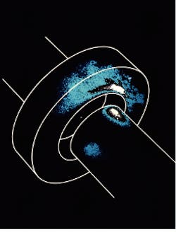
Various containment structures have been developed to enable the isolation of single or limited numbers of atomic ions for various applications. At NIST (Boulder, CO), the Max-Planck Institut (Garching, Germany), and elsewhere, scientists are investigating laser-cooled ion traps for potential use as ultra-accurate frequency and time standards and in other applications, because oscillations from single isolated atoms or ions, unperturbed by interactions with adjacent atoms, are expected to have greater spectral purity (see photo, p. S21). The Mepsicron created this image of photon emission from a single mercury ion located in a Paul-type ring ion trap. A UV laser has been used to cool the single ion in the tra¥with Doppler techniques; the 192-nm (dee¥UV) fluorescence signal detected by the Mepsicron imager camera (equipped with a lens) enables localization of the ion within the tra¥and, in principle, tracking of the ion motion with time.
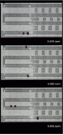
Weak hot-electron-generated optical photon luminescence occurs when CMOS devices switch on and off. A new technique promises to give IC designers an optical, noninvasive method of testing microcircuits under actual operating conditions. Devices can be spatially located in the microcircuit, and device timing can be determined by time-resolved imaging at the sub-100-ps level. Figure 4 shows time slices (actually images of a time-resolved "movie") of the optical emission from a CMOS ring oscillator. Each time-slice image corresponds to emission during a 136-ps time window out of the full 8.5-ns period of the 47-stage ring of inverters, which exhibit a stage-to-stage delay of 90 ps (in a full cycle of the ring, the pulse circulates twice). The NMOS FET of each inverter emits an ultralow-level light pulse during switching. The optical emission is shown in pseudo-color in fig. 4, while the background is an overlaid black and white microphotograph of the physical device to enable identification of the actual devices in the integrated circuit. The ring oscillator is the left half of the circuit, while the right half is a five-stage binary countdown of the ring output. Image size is about 160 ¥ 320 µm.
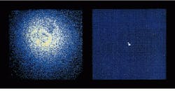
A significant advantage of space-based astronomical observations is the absence of the intervening atmosphere of the Earth. For ground-based astronomy, atmospheric turbulence normally sets a limit on the achievable resolution that can be realized, for example, to separate the images of closely spaced stars. Figure 5 shows the results of using a single-photon-counting imager, with integration times of 15 ms or less (less than the atmospheric turbulence time constant), to acquire a series of "snapshots" that were then mathematically post-processed to produce the clear, distinct "double-star" image. o
Mepsicron captures photon emission from a single mercury ion in a Paul-type ring ion trap. Ion is small emission point in center of trap.
FIGURE 1. Mepsicron imager replicates an image photon by photon on the microchannel plate (MCP) surface.
FIGURE 2. Mepsicron imager determines coordinates of imaged photons and transfers them to a PC-based display and analysis system.
FIGURE 3. Time-resolved photoluminescence mapping of emission from an AlAs/GaAs quantum-well structure shows spatial progress of plasmas through time; image-collection intervals were 0-6 ns (a), 19-25 ns (b), 38-44 ns (c), and 63-69 ns (d).
FIGURE 4. Time-resolved imaging at sub-100-ps level enables device timing measurements in complex large-scale CMOS integrated microcircuits.
FIGURE 5. Image of binary star a Hercules is rendered free of atmospheric turbulence by single- photon-counting imager and mathematical post-processing of image data.
