Thermal scanning reveals early osteoarthritis
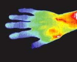
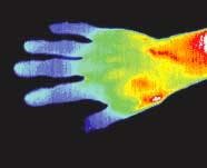
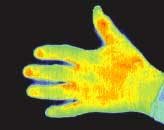
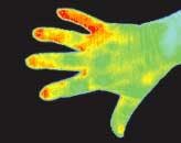
In osteoarthritis, the cartilage in one or more skeletal joints progressively breaks down. When in a knee or hip, the condition can thwart mobility; when in the hands, it can interfere with many of life's small attainments, from playing a guitar to opening a carton of milk. Traditionally, diagnosis is based on symptoms and confirmed with x-ray images. To augment this approach, researchers at Duke University Medical Center (Durham, NC) and the University of North Carolina at Chapel Hill are using thermal imaging of finger joints to produce information on the inflammatory phases of hand osteoarthritis (see figure).1
Virginia Kraus, an assistant professor of medicine and one of the researchers, notes that—although x-rays remain the standard clinical technique for diagnosing osteoarthritis—thermal scanning holds promise for detecting osteoarthritis in the first stage of the disease, before joint changes become apparent on x-rays and before symptoms such as pain and joint enlargement appear.
The study showed that finger joints in the first stage of osteoarthritis are warmer than average—a sign of inflammation. In contrast, x-rays of fingers at this early stage of the disease produce inconclusive findings, says Kraus. The temperature scans also showed that as osteoarthritis symptoms increased in severity, the joints tended to cool. Analysis showed that the progressively cooler joint temperatures correlate with increasing disease severity revealed in x-rays of the same joints, notes Kraus.
The IR-imaging system used in the experiment is produced by Compix (Lake Oswego, OR); the system's thermal-IR camera relies on 2-D scanning and a thermoelectrically cooled lead-selenide detector, is sensitive to the 3- to 5-µm wavelength band, and captures up to a 244 × 193-pixel image. The instrument has a sensitivity of 0.1°C—necessary to sense the small temperature changes induced by osteoarthritis.
Experimental findings
Ninety-one subjects were recruited for the experiment, with the selection being independent of osteoarthritis hand symptoms. Subjects were excluded, however, if they had a history of gout, psoriasis, lupus erythematosus, Raynaud's phenomenon (a condition that affects blood vessels in the extremities), or rheumatoid arthritis (which attacks the linings of the joints, rather than the cartilage).
In the thermal-IR hand images, the researchers analyzed three joints on each finger of the subjects, excluding thumbs. In addition to absolute temperature of the hands, relative temperature measurements were taken, with a spot on the wrist being the reference region. X-rays and ordinary photographs were also taken of the subjects' hands.
The researchers observed significant temperature differences between osteoarthritis-free and osteoarthritis-affected joints. The researchers relied on the Kellgren-Lawrence grading system to categorize the joints by x-rays: Grade KL0 is a normal joint, Grade KL1 is a joint with a small bony growth, or osteophyte, of doubtful significance. The scale stops at KL4, which marks severe joint-space narrowing and large osteophytes.
The study showed that joints meeting the KL1 criteria were significantly warmer than KL0 joints, while KL2 through KL4 joints were colder than KL0 joints. These results support the idea that the earliest phase of hand osteoarthritis represents an inflammatory phase of the disease, says Kraus. However, the drop in temperature as the disease worsens is an enigma. "It's unclear if it's from disuse or lack of blood supply, but temperature somehow diminishes as bony enlargements grow," says Kraus.
REFERENCE
- G. Varjú et al., Rheumatology Advance Access (May 4, 2004) and Rheumatology 43, 915, (2004).


