Photomultiplier tubes offer high-end sensitivity
When researchers and instrument designers need the highest level of sensitivity in photon detection, up to counting individual photons, they generally turn to photomultiplier tubes (PMTs). Among the oldest detector technologies (first developed in the mid-1930s), PMTs use vacuum rather than solid-state techniques to multiply electrons generated by photons. They are widely used in research activities ranging from biomedicine to astronomy as well as in medical diagnostic technology, environmental monitoring, industrial quality control, and aerospace.
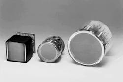
In the past few years, the development of position-sensitive PMTs has opened up the possibility of overcoming long-standing limits on spatial resolution and increasing the versatility of PMT technology. In particular, there is considerable effort in the research community to apply position-sensitive PMTs to advanced medical diagnostic and research tools such as positron emission tomography (PET, see photo at top of this page).
In all PMTs the basic principles of operation are the same. Light passes through an input window and strikes a photocathode, leading to the emission of photoelectrons into an evacuated tube. The electrons are accelerated and focused by an accelerating electrode and strike a metal dynode (see Laser Focus World, May 1996, p. 96). The multiplication takes place at the dynode: each primary electron causes the emission of several (usually around ten) secondary electrons. These are then accelerated again toward another dynode, and the process is repeated until overall multiplication factors of up to millions are achieved.
The electrons from the last dynode are collected at the anode and leave the PMT for processing by external measurement circuits. Generally, the total potential between the photocathode and anode is about 1-2 kV, and, with 10 dynodes, the drop between each dynode is 100-200 V. This differential is enough to yield good multiplication and fast response times, on the order of nanoseconds.
By selecting the appropriate photocathode material, PMTs can be made sensitive to a range of spectral windows. Cesium iodide is used for ultraviolet (UV) wavelengths from 0.115 to 0.2 µm, Sb-Cs and bi-alkali (Sb-Rb-Cs, Sb-K-Cs) are used for the visible range, and Sb-Na-K-Cs and GaAs extend from 0.3 to near-infrared (IR) at 0.85 µm. For further into the IR range, up to 1.2 µm, Ag-O-Cs and InGaAs are available, and these materials also have sensitivity extending down to 0.3 µm.
The window material also affects the range of spectral sensitivity. Borosilicate glass is the most common window material; it has a UV cutoff of 0.3 µm. For shorter wavelengths, magnesium fluoride, sapphire, and synthetic silica are used, with UV cutoffs of 0.115, 0.15, and 0.16 µm, respectively.
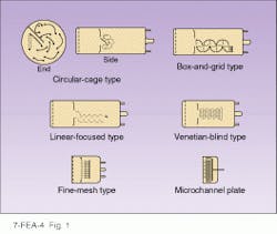
There are a number of different dynode arrangements. The most common are the circular cage, in which the electrons are bounced around in a circular pattern, and the box and grid, in which the dynodes are a series of semicircles set in a line (see Fig. 1). Another design is the venetian blind, in which each dynode consists of a series of metal slats. More recently, the fine-mesh dynode has been developed for use in position-sensitive PMTs, in which position information is preserved as the electrons pass from one grid-like dynode to the next.One dynode arrangement that has become increasingly important in the past decade is the microchannel plate (MCP). In this configuration, instead of having separate dynodes in a series, each small glass capillary channel serves as a multiple dynode, because the electrons keep bouncing back and forth off the channel walls, causing multiplication at each bounce. Each channel has an internal diameter of 6 to 20 µm, and many thousands of these are arranged in an array.
The MCPs considerably shorten the path traveled by each electron, so response times are improved to hundreds of picoseconds. In addition, MCPs address a major weakness of other PMTs—sensitivity to magnetic fields. In conventional PMTs, such fields can distort electron paths and reduce gain. But in MCPs, the electron-path details do not matter because the electrons are confined to the channels, and a degree of insensitivity to magnetic fields is achieved. Fields up to 20 kG parallel to the channels can be tolerated, although only about 700 G across the axis is allowed.
Photomultiplier tubes operate in two distinct modes, depending on the intensity of illumination. In extremely low light levels, the time between individual photons is greater than the device response time, so each photon is counted as a single pulse. As illumination increases, the pulses merge together into a time-varying analog signal. These two modes naturally require different circuitry for measurement. In the analog case, current is measured and averaged, while for photon counting, pulse-height analyzers count all pulses over a given threshold.
Scintillators and position-sensitive PMTs
By themselves, PMTs can detect radiation into the UV range. Their utility, however, is enormously widened by linking them to scintillators, allowing radiation detection far into the gamma-ray range. When radiation enters a scintillator, it produces a brief flash of fluorescence. This fluorescence is at a longer wavelength, and it can then be detected by a PMT. Common scintillator crystals used with PMTs include barium fluoride (BaF2), bismuth germania oxide (Bi4Ge3O12 or BGO), and cadmium tungstate (CdWO4).
A major application of such scintillator-PMT combinations is in the medical diagnostic technology of positron emission tomography. In this technique, radioactively labeled substances concentrate where activities of interest, such as oxidation, are taking place, allowing them to be studied in real time. The radionuclides emit positrons (the positively charged antimatter particles corresponding to electrons) that rapidly collide with and annihilate electrons, producing pairs of oppositely directed gamma rays. A ring of scintillator-PMT detectors record the gamma rays. Coincidence counting circuits define the line along which the gamma rays emerge, and, by mapping many such scintillations, a three-dimensional map of the radionuclide concentration can be built up with a resolution of about 2.1 mm. Alternatively, the time of flight to each scintillator can be measured, giving the distance along the line connecting the detectors.
For these and other applications, improvements in spatial resolution are limited by the physical size of the PMTs. As a result, there has been an intense development effort to create ways in which the position of photons on the face of the PMT can be detected and measured, thus greatly improving resolution. In PET scanning, in particular, the use of animals in research that have brains and organs much smaller than in humans has put a premium on such advances in resolution.
A number of approaches to position-sensitive PMTs have now been proposed and marketed. One approach is multianode MCPs. In these devices, an anode array, such as a 10 × 10 matrix, is located behind an MCP array. Be cause the MCP preserves position information, the anode array can pick it up accurately with a resolution of around 1.5 mm.
Most interest, however, has focused on two other approaches that use fine-mesh dynodes. In one design, a similar multianode array is used, with somewhat coarser resolution of a few millimeters. The fine mesh, which is actually a series of meshes, keeps the electrons localized as they are multiplied, because each electron travels only a short distance before hitting the next layer of mesh.
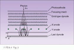
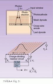
The second, more-recent design uses a coarser mesh or grid dynode, but adds a focusing mesh between each grid dynode (see Fig. 2). The magnetic fields created by the wires in the focusing mesh act as electron lenses, which causes the electrons to curve back toward their original positions and further limits the spread of the secondary electrons from the position of the primary electron.The beauty of the cross-wire grid is that it allows accurate determination of the peak or center of gravity of the broad spot created by an electron shower. While the shower may be 4 mm across at the base, the peak of the shower and thus the location of the primary electrons is measured to an accuracy of a few tenths of a millimeter. The actual accuracy depends on how many photons are generated by a given scintillation at a given spot in the crystal. The trick is done with amazing simplicity: the x coordinate of the originated spot is found by dividing the charge from one end of the X set by the sum of the charge from both ends, and the y coordinate the same way.
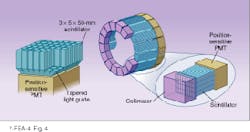
Researchers at Hamamatsu Photonics (Bridgewater, NJ), a developer of position-sensitive PMTs, have used cross-wire PMTs with arrays of BGO scintillators to show that PET scanners can be built with resolutions of 2-3 mm, good enough for detailed experimental studies in small animals (see Fig. 4).Applications of PMTs
In addition to applications such as PET scanners, PMTs are used widely in biomedicine. One of the most sensitive methods of analysis in medicine is radioimmunoassay. In this technique, radioactively labeled antibodies are introduced to measure the presence of target antigens. When the antibodies bind to the antigens, they can be precipitated out and the strength of the their radiation used to measure extremely small amounts. Samples are inserted into shielded cavities with scintillators and PMTs that carry out the measurement.
In addition, PMTs are central to the workhorse instrument of the genetic-engineering revolution, the DNA sequencer (see Laser Focus World, Oct. 1995, p. 135). Determining the sequence of bases on a given section of DNA is the first step in mapping a portion of DNA and thus being able to study and eventually manipulate it. In the sequencing process, DNA from a cell is injected onto a gel electrophoresis plate, along with a fluorescent label that binds to the DNA. Under the effect of an electric field, the DNA separates out into different segments based on size and charge. As the DNA passes under laser illumination, the fluorescence label is excited. The light is filtered and passed to PMTs, which record it. Computer analysis of the position at which the fluorescence was emitted enables a determination of the DNA sequence.
In industry, PMTs also show up in much more mundane uses. For example, color scanners used in four-color reproduction processes have banks of PMTs behind color filters to provide precise coding of color information. Oil-well drilling is another use of ruggedized PMTs, and a host of environmental and monitoring instruments have PMT detectors.
At the other extreme, PMTs are being deployed in the most sensitive and exacting astronomical and high-energy physics experiments. For example, in the 1980s, physicists developed speculative theories that indicated that protons, given several hundred billion years, would decay.
Neutrino detection
To test this theory, a 50,000-ton tank of purified water in Kamiokande, Japan, was surrounded with 10,000 20-in. PMTs. The theory was that a decaying proton would emit high-energy particles traveling faster than the speed of light in water (which is slower than the speed of light in a vacuum). In this situation, Cerenkov light is emitted by the particle, just as a supersonic aircraft emits a sonic boom. The light would be detected by the huge PMT bank.
So far, no proton decays have been found (protons, it seems, last forever), but the experiment successfully detected neutrinos, highly penetrating particles from a nearby supernova. The neutrinos, by interacting with hydrogen nuclei, could change a proton to a neutron, causing the emission of a high-speed electron, which again would be detected by the PMT array. Similar and larger experiments are now used to study the mysterious deficit of neutrinos from the sun, which has long puzzled astronomers. (The sun produces only about one-third as many neutrinos as theory predicts.) Quite possibly in the next few years, such huge PMT arrays floating in the ocean (the Deep Underwater Muon and Neutrino Detector project in Hawaii, see Laser Focus World, Dec. 1995, p. 100) will bring back news of fundamental new discoveries about the universe.
About the Author
Eric J. Lerner
Contributing Editor, Laser Focus World
Eric J. Lerner is a contributing editor for Laser Focus World.
