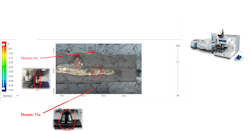
Jacopo Tintoretto, a 16th century Italian painter, created large-scale narrative paintings featuring dramatic lighting and gestures. Among his most notable are paintings that adorn the Scuola Grande di San Rocco in building in Venice—founded in the late-15th century as a confraternity (a Christian voluntary association that promotes charity); namely the Sala dell’Albergo, where meetings of Scuola’s governing body were once held (see Fig. 1). They canvas three main ceilings in the Sala Capitolare room and narrate key moments in the journey of the Israelites, including Christ’s crucifixion and the Old and New Testaments.
These irreplaceable centuries-old paintings are steeped in rich history, according to researchers at the Ca’Foscari University Venice, so conservation has been a priority for several years. The efforts began in 2018 with curators at the Scuola Grande and the Venice in Peril Fund, a U.K.-based charity that raises funds for the conservation of at-risk buildings, monuments, and works of art across Venice and its islands.
Now, they’re getting a boost from Thermo Fisher Scientific’s Nicolet RaptIR Fourier transform infrared (FTIR) microscope (see Fig. 2).Analyzing artwork
The FTIR microscope offers precision and agility to help streamline sample analysis by quickly generating actionable results, including the ability to view the intricacies of a sample, making it well suited for art restoration. The technology is highly adaptable, according to Michael S. Bradley, senior manager of FTIR spectrometers and FTIR microscope products at Thermo Fisher. It uses both visual light, which shows colors and layers, and infrared (IR) light. The IR beam focuses onto the sample, which is located on a moving stage, down to a 5 µm spot size. The IR light permits the identification of microscopic components in a sample.
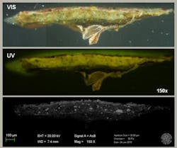
The Ca’Foscari team took small samples and used the microscope’s IR component to analyze them, says Eleonora Balliana, a researcher with the Department of Environmental Sciences, Informatics, and Statistics at Ca Foscari. They also used a cross-section to perform different types of analysis without compromising the sample (see Fig. 3).“The concern is always that the sample will need to be too compressed or handled too aggressively and it will therefore sustain damage,” she says. “The RaptIR does not damage the samples, which is especially important in circumstances where the sample simply cannot be replaced.”
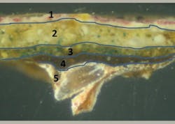
Researchers have been able to confirm the composition of the sometimes-unusual materials Tintoretto used to compose these historic paintings. “The ability to work directly on a cross-section enabled us to better investigate the possible differences in terms of pigments and the binders among the paint layers,” Balliana says (see Fig. 4).With visual spatial resolution at 1 µm and IR spatial resolution at better than 5 µm, the FTIR microscope allows the researchers to acquire higher-quality, higher-resolution spectra of the samples. Diffraction-limited objectives and auto-illumination have offered better visual and IR images of the paintings, according to Barbara Bravo, an FTIR and Raman application specialist with Thermo Fisher. The microscope also makes it possible to directly analyze the works without any sample preparation—this is key to preserving irreplaceable artwork.
“Ultimately, the quality of the analysis is much improved with an FTIR microscope in contrast to the analysis results provided by alternative technologies,” Balliana says.
The FTIR microscope technology also provides fast results, with mapping of up to 10 spectra/second. “Any amount of time saved while searching for the solution makes a world of difference in terms of delivering results in a timely and accurate manner,” says Bravo, adding that such technology propels detailed research forward, providing enhanced images that are better defined than those obtained via a standard or conventional microscope.
Understanding the artist
Using modern microspectroscopy to achieve higher visual acuity and IR quality has been at the center of the research team’s goals to unearth detailed information about Tintoretto’s use of oils as binders and pigment types, including the presence of the earth-based pigments and animal proteins.
The FTIR microscope has revealed the individual layers of the works on the canvas, providing the researchers with a deeper insight into Tintoretto as well as the materials he chose to work with and why. The analysis revealed the various layers of the paintings, starting from the top visible section (the pictorial portion) and into the first layer of oil against the canvas that helped the materials spread evenly over it. The team discovered an animal glue that sealed the pores of the canvas, preventing the paint from being absorbed into the porous surface.
Tintoretto manipulated the various nonconventional materials he used to work fast and cover a larger area of canvas without excessive absorption, which ultimately saved money on painting supplies. This deeper understanding of the paint chemistry the microscope provides has also offered valuable knowledge about how to clean, treat, and better preserve such irreplaceable works of art.
Next steps
Balliana says the team plans to expand its use of FTIR microscopy to other art samples and present the data together with additional forms of analysis—noninvasive imaging, gas chromatography-mass spectrometry, scanning electron microscopy, and energy dispersive x-ray spectroscopy—to create a dedicated publication about Tintoretto’s paintings. Despite great interest in the artist’s techniques and masterpieces, few publications are related to the Sala Capitolare space that features his works.
“The collected results will help expand the existing knowledge available about Tintoretto’s painting techniques in comparison to other artworks present in the Scuola Grande and around Venice. This will also provide important data about the environmental conditions present where the artwork is housed; in this case, the Scuola Grande,” Balliana says. The data provides expanded knowledge of the paint materials, as well, ultimately offering potential options for crucial subsequent conservation interventions in relation to previous interventions.
About the Author
Justine Murphy
Multimedia Director, Digital Infrastructure
Justine Murphy is the multimedia director for Endeavor Business Media's Digital Infrastructure Group. She is a multiple award-winning writer and editor with more 20 years of experience in newspaper publishing as well as public relations, marketing, and communications. For nearly 10 years, she has covered all facets of the optics and photonics industry as an editor, writer, web news anchor, and podcast host for an internationally reaching magazine publishing company. Her work has earned accolades from the New England Press Association as well as the SIIA/Jesse H. Neal Awards. She received a B.A. from the Massachusetts College of Liberal Arts.


