New imaging technique aids photothermal therapies
With the potential to enhance photothermal therapy, real-time, more accurate temperature detection could help doctors and clinicians diagnose skin cancers earlier and treat them quicker and ideally more effectively.
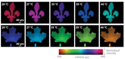
Researchers at the Quebec (Canada)-based Institut National de la Recherche Scientifique (INRS) have developed single-shot photoluminescence lifetime imaging thermometry (SPLIT), which can easily measure temperature in 2D, and with no contact.
The new imaging technique is based on the luminescence of nanoparticles doped with rare-earth ions. According to researcher Fiorenzo Vetrone, a professor at INRS, the nanoparticles are considered nanothermometers, as “their luminescent properties change with the temperature of the environment;” they are biocompatible as well.
“Compared to existing thermometry techniques, SPLIT is faster and has higher resolution,” says Jinyang Liang, a professor at INRS and a lead researcher in ultrafast imaging. “This allows a more accurate temperature sensing with both an advanced and economical solution.”
The new technique eliminates the time-consuming process of imaging the luminescence point by point. Instead, the SPLIT technology touts an ultra-high-speed camera that can track the speed of the luminescence decay of the nanoparticles at each spatial point.
“Our camera is different from a common one, where each click gives one image. Our camera works by capturing all of the images of a dynamic event into one snapshot. Then we reconstruct them, one by one,” says Xianglei Liu, a Ph.D. student at INRS and lead author of the study, adding that from there, SPLIT can sense the temperature “by checking how fast the emitted light fades out. Since it is in real time, SPLIT can follow the phenomenon as it happens.”
SPLIT has the potential to detect skin cancers such as melanoma, in particular, more quickly. The researchers note that existing imaging techniques are invasive, they produce lower-resolution images, and are less accurate, all of which “leads to a large number of unnecessary biopsies” and other related procedures. The new technology could also be used to monitor and better control aspects of treatment such as light doses.
About the Author
Justine Murphy
Multimedia Director, Digital Infrastructure
Justine Murphy is the multimedia director for Endeavor Business Media's Digital Infrastructure Group. She is a multiple award-winning writer and editor with more 20 years of experience in newspaper publishing as well as public relations, marketing, and communications. For nearly 10 years, she has covered all facets of the optics and photonics industry as an editor, writer, web news anchor, and podcast host for an internationally reaching magazine publishing company. Her work has earned accolades from the New England Press Association as well as the SIIA/Jesse H. Neal Awards. She received a B.A. from the Massachusetts College of Liberal Arts.

