Raman detection refinements offer detailed chemical imaging
When a light beam passes through a material, some photons in the beam can gain or lose energy interacting with certain molecular vibrations; this is referred to as the Raman effect. The transmitted spectrum is informative, but signal intensity can be 1012 times weaker than the incoming beam.
Coherent Raman scattering amplifies the signal, illuminating a sample with two beams, a pump, and a Stokes beam. A signal is generated when the energy difference between the two beams matches a vibrational state in the sample molecules. This principle supports both coherent anti-Stokes Raman spectroscopy (CARS), which measures the blue-shifted beam, and stimulated Raman spectroscopy (SRS), which measures the stimulated Raman loss (SRL) in the pump beam and/or the stimulated Raman gain (SRG) in the Stokes beam.
Professors Giulio Cerullo and Dario Polli and postdoc Alejandro De la Cadena in the Department of Physics at Politecnico di Milano (Italy) discussed several of their approaches to broadband coherent SRS imaging at SPIE’s 2021 Photonics West Conference.1
Adding power to spectroscopic analysis
CARS and SRS create signals about a million times stronger than spontaneous Raman scattering, but they’re still about a million times smaller than the incident beam. CARS signals are wavelength-shifted, but can be lost in a background of nonresonant emission. SRS measures the pump and/or Stokes beams—these variations are very small, about 10-5 of the incident beam intensity. The challenge for SRS is to find a method of isolating small variations riding on a large baseline intensity.
The latest advance by Cerullo’s team is a multichannel configuration that allows pixel-by-pixel hyperspectral analysis on a subcellular scale. The measurement principle is straightforward: simultaneously illuminate a sample with a narrowband Stokes pulse and a broadband pump pulse. Where the energy difference between the Stokes pulse and a specific wavelength in the pump pulse matches a vibrational transition energy in the sample, the pump intensity at that wavelength will be diminished. Comparing the transmitted pump intensity when the Stokes pulse is on and when it’s off provides a spectrum at that location. Simple in concept, but not necessarily simple in implementation.
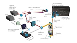
Cerullo’s team selected a ytterbium-based fiber laser as the core of the system because it’s a cost-effective, compact source emitting 140 fs, 1040 nm pulses at 80 MHz. However, fiber lasers have intensity noise that can swamp SRG or SRL signals. To be insensitive to noise while sensitive to small signals, the researchers invented an inline balanced detection measurement architecture.2
Attention to detail
The research team split the laser output, leaving part for the narrowband Stokes pulse that is modulated, while another portion generates a broadband pump signal from a purpose-built optical parametric oscillator.3 The broadband pulse is sent to a prism compressor, which introduces dispersion tuned to compensate for the opposite sign dispersion in a downstream high-numerical-aperture microscope objective; this illuminates the sample.
To suppress intensity noise, the broadband pump pulse is sent through a birefringent crystal, creating about a 5 ps delay between two orthogonal pulses with identical intensity profiles. The leading broadband pulse is transmitted through the sample in the absence of the Stokes pulse, providing a reference intensity. When the trailing broadband pulse and synchronous narrowband Stokes pulse transmit through the sample together, the pump beam is modified by SRL. The baseline pump pulse and the Raman-modified version are routed to balanced photodetectors, sensitive only to intensity difference and not the common noise. When that difference signal is sent to a lock-in amplifier matched to the Stokes modulation frequency, the output represents the part of the difference associated with the presence of the Stokes beam—that is, it directly measures the SRL.
That quantifies total SRL over the entire spectrum. The final step is to replace the single detector pair with dispersive optics and two matched linear arrays of photodetectors. Each matched pair then directly measures the loss at that specific wavelength range, providing a spectral signature to identify constituent molecules. By scanning the incident beams across the field of a microscope objective, the method creates a map of the sample’s chemical distribution at microscopic scale. Previously, Cerullo’s team developed a four-band detector with balanced detection implemented in custom CMOS circuitry.4 They have now extended that work and will soon publish results from a 32-band implementation with spectral resolution of better than 8 cm-1 per pixel, providing pixel-by-pixel hyperspectral vibrational spectra.
After several years of development, Cerullo says, “it was gratifying to finally see the Raman images come out of the instrument.” He believes broadband SRS microscopy will open up opportunities in cell biology, medical diagnostics, and other fields, as “it enables noninvasive imaging of chemical composition of substances, cells, and tissues with unprecedented speed and spatial resolution.” Cerullo has founded Cambridge Raman Imaging (cambridgeramanimaging.com) to commercialize the technology.
REFERENCES
1. A. De la Cadena et al., Proc. SPIE, 11656, (2021); https://doi.org/10.1117/12.2578587.
2. F. Crisafi et al., Sci. Rep., 7, 10745 (2017); https://doi.org/10.1038/s41598-017-09839-1.
3. N. Coluccelli et al., Opt. Lett., 42, 4545–4548 (2017); https://doi.org/10.1364/ol.42.004545.
4. G. Sciortino et al., IEEE J. Solid-State Circuits, 56, 1859–1870 (2021); https://doi.org/10.1109/jssc.2020.3046484.
About the Author

Richard Gaughan
Contributing Writer, BioOptics World
Richard Gaughan is the Owner of Mountain Optical Systems and a contributing writer for BioOptics World.