Microdiamonds boost both optical imaging, MRI
Faster, deeper imaging is possible with the help of microscopic diamond tracers that researchers at University of California, Berkeley (UC Berkeley) have demonstrated to provide information via optical fluorescence, as well as MRI, simultaneously.
“There is a lot of information you can get in combination because the two modes are better than the sum of their parts,” says Ashok Ajoy, an assistant professor of chemistry at UC Berkeley and co-author of the study. “This opens up many possibilities, where you can accelerate the imaging of these diamond tracers in a medium by several orders of magnitude.”
The technique has the potential for scientists to obtain higher-quality images faster and up to a centimeter below surface tissue. This is about 10X deeper than traditional light-based techniques, according to the researchers. Traditional light microscopes allow researchers to view submicron-resolution structures inside cells or tissue, but “only as deep as the millimeter or so that light can penetrate without scattering.”
MRI imaging experiences similar challenges, as it can only provide low-resolution images, and about 1000X worse than what light can do.
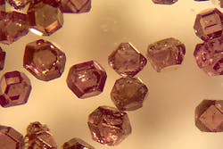
In the UC Berkeley study, the promising technique uses a newer biological tracer—microdiamond—that has had some of its carbon atoms eliminated and replaced by nitrogen. The researchers say this leaves empty spots in the crystal, called nitrogen vacancies, which fluoresce when hit by laser light. Further, carbon-13, which occurs naturally in diamond particles at about 1% concentration and has the potential for additional enrichment via replacing dominant carbon atoms, was exploited. According to the researchers, carbon-13 nuclei are more readily polarized by “nearby spin-polarized vacancy centers, which become polarized at the same time they fluoresce after being illuminated with a laser.” This polarized nuclei ultimately produces a stronger signal for MRI.
The team found that the hyperpolarized diamonds can be detected optically, thanks to the fluorescent nitrogen vacancy centers, and also at radio frequencies (for MRI) because of the spin-polarized carbon-13. This allows simultaneous imaging by two of the best techniques available, with particular benefit when looking deep inside tissues that scatter visible light.
“Optical imaging suffers greatly when you go in deep tissue. Even beyond 1 mm, you get a lot of optical scattering. This is a major problem,” Ajoy says. “The advantage [discovered in this study] is that the imaging can be done in radio frequencies and optical light using the same diamond tracer. The same version of MRI that you use for imaging inside people can be used for imaging these diamond particles, even when the optical fluorescence signature is completely scattered out.”
Ajoy and his team note that this technique could be ideal for studying cells and tissue outside the body, probing blood or other fluids for chemical markers of disease, or for physiological studies in animals. Reference: X. Lv et al., Proc. Nat. Acad. Sci. (2021); doi.org/10.1073/pnas.2023579118.
About the Author
Justine Murphy
Multimedia Director, Digital Infrastructure
Justine Murphy is the multimedia director for Endeavor Business Media's Digital Infrastructure Group. She is a multiple award-winning writer and editor with more 20 years of experience in newspaper publishing as well as public relations, marketing, and communications. For nearly 10 years, she has covered all facets of the optics and photonics industry as an editor, writer, web news anchor, and podcast host for an internationally reaching magazine publishing company. Her work has earned accolades from the New England Press Association as well as the SIIA/Jesse H. Neal Awards. She received a B.A. from the Massachusetts College of Liberal Arts.

