Lithium fluoride fluorescent crystals track high-energy heavy ions such as iron
Physicists from the Institute of Nuclear Physics of the Polish Academy of Sciences (IFJ PAN; Kraków, Poland) have just demonstrated that lithium fluoride (LiF) crystals, which previously have been used to register the tracks of nuclear particles such as alpha particles (helium nuclei), are also ideal for detecting tracks of high-energy ions of elements as heavy as iron, completely stripped of electrons.1
"Lithium fluoride track detectors are simply crystals," says Paweł Bilski, a researcher at IFJ PAN. "Unlike detection devices that monitor near-real time tracks of particles, they are passive detectors. In other words, they work like photographic film. Once crystals are exposed to radiation, we need to use a fluorescence microscope to find out what tracks we have recorded."
Fluorescent nuclear track detectors have been known for about a decade. So far, they have been made only from appropriately doped aluminum oxide (Al2O3) crystals in which, under the influence of radiation, permanent color centers are created. Such centers, when excited by light of an appropriate wavelength, emit photons (with lower energies) that make it possible to see the track of a particle under a microscope. In the case of LiF, the excitation is carried out with blue light and the emission of photons takes place in the red range.
"Detectors with doped aluminum oxide require an expensive confocal microscope with a laser beam and scanning," says Bilski. "Tracks in lithium fluoride crystals can be seen with a much cheaper, standard fluorescent microscope. Tracks recorded in crystals very accurately reproduce the path of a particle. Other detectors, such as the well-known Wilson chamber, usually widen the track. In the case of LiF crystals, the resolution is restricted only by the diffraction limit."
Personal dosimetry
While the impossibility of observing tracks of particles in near real time is difficult to call an advantage, it does not always have to be a disadvantage. For example, in personal dosimetry, detectors are needed to determine the dose of radiation to which the user has been exposed. These devices must be small and easy to use. Millimeter-sized crystalline LiF plates meet this requirement well. This is one of the reasons why these crystals, grown by Czochralski method at IFJ PAN, can now be found in the European Columbus module of the International Space Station, among many other types of passive detectors. Replaced every six months within the DOSIS 3D experiment, the detectors make it possible to determine the spatial distribution of the radiation dose within the station and its variability over time.
During the latest research, crystalline lithium fluoride plates were exposed to high-energy ions. The irradiation was carried out in the HIMAC accelerator in the Japanese city of Chiba. During the bombardment with various ion beams, the energies of particles ranged from 150 MeV/nucleon in the case of 4He helium ions to 500 MeV/nucleon in the case of 56Fe iron ions. The detectors were also irradiated at with 12C carbon ions, 20Ne neon, and 28Si silicon beams.
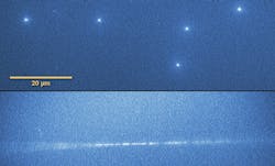
"In the crystal plates placed perpendicularly to the ion beam, we observed practically point sources of light of a size on the border of the optical resolution of a microscope," says Bilski. "These were the places where the high-energy ion pierced the crystal. As part of the tests, some of the plates were also placed parallel to the beam. The probability of registering a track was then lower, but when it did happen, a long fragment of the track of the particle was 'imprinted' in the crystal."
Personal neutron detectors
In addition, LiF detectors, especially those enriched with the 6Li isotope, may effectively track low-energy neutrons (and maybe higher-energy as well). If future studies confirm this assumption, it will be possible to construct personal neutron dosimeters. The small size of LiF crystals would also allow for interesting technical applications that are technologically inaccessible today. LiF track detectors could be used, for example, to study secondary particles formed around the primary proton beam produced by accelerators used in medicine to fight cancer.
Source: https://press.ifj.edu.pl/en/news/2019/08/19/
REFERENCE:
1. Paweł Bilski et al., Journal of Luminescence (2019); https://doi.org/10.1016/j.jlumin.2019.05.007.
About the Author
John Wallace
Senior Technical Editor (1998-2022)
John Wallace was with Laser Focus World for nearly 25 years, retiring in late June 2022. He obtained a bachelor's degree in mechanical engineering and physics at Rutgers University and a master's in optical engineering at the University of Rochester. Before becoming an editor, John worked as an engineer at RCA, Exxon, Eastman Kodak, and GCA Corporation.

