Mid-IR upconversion imaging promises to speed diagnosis
Because it enables label-free, high-specificity detection of chemical signatures, mid-infrared (mid-IR) spectral imaging is well suited for biological specimen assessment. But a lack of affordable light sources and sufficiently sensitive detectors has hindered acceptance of mid-IR methods—in fact, existing camera technology is most sensitive in the near-infrared (near-IR) region. Now, a system developed by an international research collaborative bridges this gap.
The team used nonlinear frequency conversion (upconversion), a process in which energy is added to a photon to change its wavelength. But instead of applying it to change the wavelength output of a laser, which is often done, researchers from DTU Fotonik (Oskild, Denmark), the Barcelona Institute of Science and Technology and Instituto Catalana de Recerca (both in Barcelona, Spain), the University of Exeter (Exeter, England), and the Gloucestershire Hospitals NHS Foundation Trust (Cheltenham, England) applied it to shift an entire mid-IR image into the near-IR range while preserving all the original spatial information.1
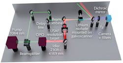
The system (see figure) incorporates a single-wavelength mid-IR light source, developed at the Institute of Photonics Sciences (ICFO), that can be tuned to different wavelengths. To generate the mid-IR light, the source also uses frequency conversion. In fact, the researchers used the same pulsed near-IR laser both to generate tunable mid-IR light and to achieve image upconversion. Compared with direct mid-IR detection, upconversion provides orders-of-magnitude greater sensitivity and speed. A synchronously pumped optical parametric oscillator (OPO) produces 20 ps pulses tunable in the 2.3–4 μm spectral range for mid-IR illumination. (While the 5–12 μm range is typically preferred for chemical specificity, the 3–4 μm range also shows relevant chemical features, and has the extra advantage of being compatible with commonly used microscope slides.)
A lithium niobate crystal serves as the birefringent phase-matched nonlinear medium for upconverting mid-IR signals to the near-IR range for fast capture by a standard CCD camera. The team demonstrated image-capture speed by tuning the laser to match the peak absorption of a gas flow and acquiring video at 40 fps.
Crystal rotation increases field of view
To increase the field of view (FoV), the researchers revived a decades-old idea: angular crystal rotation. Crystal rotation generates phase matching of concentric rings in the object plane, and thus upconverts distinct ring-shaped regions as a function of time. The team enabled rotation in single-degree increments by mounting the crystal on a galvanometer scanner and using tangential phase matching. Compared to fixed-angle configuration, such fine rotation enables an increase in the FoV by roughly a factor of five and boosts pixel count in the upconverted image by about a factor of 25. Thus, FoV is limited primarily by lens optics, just as in standard imaging configurations—and incidentally, this concept can be applied to any upconversion imaging system.
Altering crystal rotation cycle time to match the camera’s integration time allows direct acquisition of imagery with increased FoV—without postprocessing. By comparison, previous work involving monochromatic upconversion required considerable postprocessing to produce a large FoV.
The researchers demonstrated monochromatic mid-IR upconversion imaging with 2.5 ms frame acquisition. This allowed 35 μm spatial resolution within a FoV measuring 10 mm in diameter, and thus provided about 64 kpixels (spatially resolvable elements) in the object plane. Team members from Exeter University used the system to evaluate and distinguish between cancerous and healthy esophageal tissue samples. The approach, they say, generated morphology and spectral classification output that aligned well with standard histopathology imagery.
Standard histopathology relies on a labor-intensive process of staining of tissue sections with hematoxylin and eosin (H&E), followed by subjective manual assessment using an optical microscope. Histopathological mid-IR hyperspectral imaging (HSI) requires less sample preparation and provides objective information based on chemical signatures of proteins, lipids, and DNA. Because molecular composition of tissue is known to change with disease progression, the spectral data can be used for computer-assisted classification of diseases. In fact, the researchers predict that the addition of machine learning could enable faster diagnostics. Thus, the technology promises to facilitate investigation of cancer and other diseases by increasing accuracy and speeding diagnoses.
REFERENCE
1. S. Junaid et al., Optica (2019); https://doi.org/10.1364/optica.6.000702.
About the Author

Barbara Gefvert
Editor-in-Chief, BioOptics World (2008-2020)
Barbara G. Gefvert has been a science and technology editor and writer since 1987, and served as editor in chief on multiple publications, including Sensors magazine for nearly a decade.
