Raman spectroscopy helps distinguish tumors that resist radiation treatment
A team of researchers at the University of Arkansas (Fayetteville, AR), Johns Hopkins University (Baltimore, MD), and the University of Arkansas for Medical Sciences (UAMS; Little Rock, AR) has started pilot clinical studies in head and neck cancer patients to determine if Raman spectroscopy can effectively spare some patients of the toxic side effects of ineffective radiation therapy.
In using Raman spectroscopy, an optical fiber-based, noninvasive, and highly specific imaging technique, the research team discovered differences between treatment-sensitive and treatment-resistant tumors in mice after radiation. Their findings revealed statistically significant differences in lipid and collagen content that could potentially identify treatment-resistant tumors early in the therapeutic regimen.
"Identifying patients with radiation-resistant tumors prior to commencing treatment or immediately after it has begun would significantly improve response rates and help these patients avoid the toxic side effects of ineffective radiation therapy," says Narasimhan Rajaram, assistant professor of biomedical engineering at the University of Arkansas. "Our findings provide a rationale for translating these studies to patients with this as the ultimate goal."
Rajaram and Ishan Barman, assistant professor of mechanical engineering at Johns Hopkins University, used Raman spectroscopy to map biomolecular changes in human lung tumors and two different types of head and neck tumors. Their research team found that radiation-sensitive tumors had greater changes in the expression of lipids and collagen. These changes were consistent across all tumors.
These findings follow a similar study published last year, in which Rajaram and an interdisciplinary team of researchers at the University of Arkansas and UAMS used autofluoresence imaging to identify differences between the metabolic response of lung cancer cells after exposure to radiation and YC-1, a common chemotherapy drug.
Related: Fluorescence imaging helps localize lung tumors in real time
Radiation is used, along with chemotherapy, to treat the majority of patients diagnosed with lung, head, or neck cancers. While these treatments can last up to seven weeks, there are no accepted methods to determine treatment response before or during the early stages of therapy, which means some patients undergo a full treatment regimen only to be identified later as nonresponders.
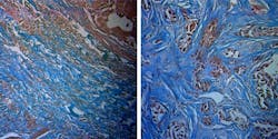
The lung cancer cells used in this study were developed by Ruud Dings, assistant professor of radiation oncology, and Robert Griffin, professor of radiation oncology at UAMS. Histological evaluations of these tumors by Matt Quick, associate professor of pathology at UAMS, were consistent with the Raman spectroscopic results.
Sina Dadgar and Paola Monterroso Diaz, graduate students in biomedical engineering, were lead authors on the study, along with Santosh Paidi, a graduate student at Johns Hopkins University.
Full details of the work appear in the journal Cancer Research.
