Raman spectroscopy method could diagnose bone diseases noninvasively
A team of researchers from University College London (UCL; London, England), the Science and Technology Facilities Council (STFC; Swindon, Wiltshire, England), and the Royal National Orthopaedic Hospital (RNOH; also in London) used a Raman spectroscopy method that could enable doctors to identify bone diseases such as osteoporosis and osteogenesis imperfecta (OI, known as brittle bone disease) without having to use invasive diagnostic methods or exposing patients to radiation associated with current x-ray techniques.
Related: FTIR spectroscopy helps predict bone density, fracture risk
This research has, for the first time, enabled detection of OI (a genetic disease) by simply scanning a patient's limbs. Until now, bone diseases have been diagnosed through x-rays, history of fractures, and other clinical symptoms and, in the case of OI, genetic testing.
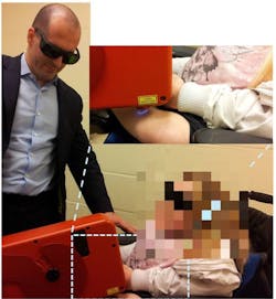
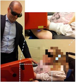
The researchers used a method known as spatially offset Raman spectroscopy (SORS) to test for the condition. The method involves shining a laser through the skin to analyze the underlying chemistry of the bone, and can reveal differences between healthy and diseased bone. The general SORS concept was developed at the STFC Central Laser Facility, based at the Research Complex in Harwell, Oxfordshire.
To obtain a set of control data, the research team carried out tests on a small bone sample taken from a 26-year-old female patient with type IV OI. This is a moderate form of the disease that affects growth and can cause bone deformity and spinal curvature.
Using conventional Raman spectroscopy, the team probed the bone and compared it to a non-diseased bone sample, establishing a notable chemical difference in its makeup.
The patient's body was then scanned extensively and noninvasively, using a laser in a custom-built SORS instrument developed by Cobalt Light Systems (Abingdon, Oxfordshire, England). For comparison, a second set of scans were carried out on a healthy female volunteer of a similar age, who does not have the disease.
“Bone is a complex material that has both mineral and protein components,” says Dr. Kevin Buckley from STFC's Central Laser Facility, one of the researchers working on this project. “Traditional x-ray methods that are used to study bone can only see the mineral, but this technique can see both components.”
The OI patient's bone sample was found to be significantly more mineralized than the non-diseased sample, and was therefore structurally brittle.
Prof. Allen Goodship of UCL's Institute of Orthopaedics and Musculoskeletal Science, who led the research, comments that the SORS method could become a routine tool that doctors can use during an annual check-up, allowing doctors to advise patients on lifestyle changes that could slow the progress of the disease further. With regular screening, SORS can monitor the effects directly, he adds.
The research was funded by a $2.6 million (£1.7 million) grant from the Engineering and Physical Sciences Research Council, with facility time and other support coming from the Science and Technology Facilities Council. Control bone samples were provided by the Vesalius Clinical Training Centre (Bristol, England).
Full details of the work appear in the journal IBMS BoneKEy; for more information, please visit http://dx.doi.org/10.1038/bonekey.2014.97.
-----
Follow us on Twitter, 'like' us on Facebook, connect with us on Google+, and join our group on LinkedIn
Subscribe now to BioOptics World magazine; it's free!

