Handheld Raman scanner could assist in complete brain tumor removal
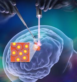
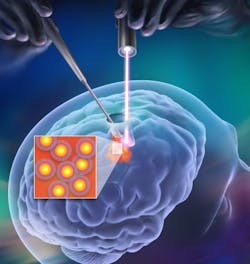
As cancerous brain tumors can grow back despite surgical attempts to remove them, scientists at the Memorial Sloan-Kettering Cancer Center (MSKCC; New York, NY) and colleagues have developed a method that relies on surface-enhanced Raman scattering (SERS) to distinguish malignant from healthy cells during surgery. In this way, fewer or no malignant cells get left behind to form new tumors.
Related: Stimulated Raman scattering could help boost accuracy of brain tumor surgery
Moritz F. Kircher, MD, Ph.D., a MSKCC radiologist who led the work, point outs that malignant brain tumors, particularly the kind known as glioblastoma multiforme (GBM), are among the toughest to beat. Surgical removal is one of the main weapons doctors have to treat brain tumors; however, there is no way to know if all cancerous cells have been removed. So, Kircher and his team used a handheld device resembling a laser pointer that, when guided by SERS nanoparticles, can identify tumors. The nanoparticles are injected the day prior to the operation and go specifically to tumor cells, and not to normal brain cells. Using a handheld Raman scanner in a mouse model that mimics human GBM, the researchers identified and removed all malignant cells in the rodents' brains.
Also, because the technique involves steps that have already made it to human testing for other purposes, the researchers conclude that it has the potential to move readily into clinical trials. Surgeons might be able to use the device in the future to treat other types of brain cancer, they say.
Full details of the team's work appear in the journal ACS Nano; for more information, please visit http://dx.doi.org/10.1021/nn503948b.
-----
Don't miss Strategies in Biophotonics, a conference and exhibition dedicated to development and commercialization of bio-optics and biophotonics technologies!
Follow us on Twitter, 'like' us on Facebook, and join our group on LinkedIn
Subscribe now to BioOptics World magazine; it's free!