Noninvasive Raman microspectroscopy technique boosts stem cell therapy

Oct. 20, 2011
Researchers at Nottingham University have developed a noninvasive Raman microspectroscopy (RMS) technique that phenotypically identifies live cardiomyocyte cells within highly heterogeneous cell populations with greater than 96% sensitivity and specificity.

In stem cell therapy, a problem that exists is controlling the excessive proliferation of cells with unwanted phenotypes after transplantation to prevent tissue overgrowth and tumor formation. And most techniques currently available for characterizing cells are invasive, therefore rendering the cells unusable. Recognizing this, researchers at Nottingham University (Nottingham, England) have developed a noninvasive Raman microspectroscopy (RMS) technique that phenotypically identifies live cardiomyocyte cells within highly heterogeneous cell populations with greater than 96% sensitivity and specificity.

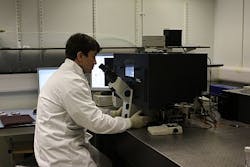
Led by Dr. Ioan Notingher, the team used a back-illuminated CCD camera (from Andor Technology) attached to a purpose-built Raman microspectrometer to record spectra from individual cells derived from micrometric regions of human embryonic stem cells (hESCs). By comparing with matching immunofluorescence images from the same cells, they showed that the Raman spectra correspond to the spatial distribution of biomolecules such as nucleic acids, proteins, lipids and carbohydrates, and that this can be used to discriminate between different cell types.

To yield the shortest possible acquisition times for the Raman spectra, the team's CCD camera enabled measurements of Raman spectra from selected positions in the cells in only 0.5 sec, says Notingher. The camera's detectors work in the 800-900 nm spectral range, thereby avoiding photodamage to the cells. And since RMS has only a minimal background signal from water, it allows repeated observations of viable cells maintained under physiological conditions, he adds.

-----

Follow us on Twitter

Subscribe now to Laser Focus World magazine; it's free!

About the Author

LFW Staff

Published since 1965, Laser Focus World—a brand and magazine for engineers, researchers, scientists, and technical professionals—provides comprehensive global coverage of optoelectronic technologies, applications, and markets. With 80,000+ qualified print subscribers in print and over a half-million annual visitors to our online content, we are the go-to source to access decision makers and stay in-the-know.

Sponsored Recommendations

On demand webinar: Meet BMF’s first hybrid resolution printer, the microArch D1025

July 26, 2024
Join us in this webinar to explore our newest product release - the microArch D1025 - our first dual-resolution printer. Learn more!

Meet the microArch D1025: Hybrid Resolution 3D Printing Technology

July 26, 2024
Meet BMF's newest release, our first dual-resolution printer for the prototyping and production of parts requiring micron-level precision.

Optical Power Meters for Diverse Applications

April 30, 2024
Bench-top single channel to multichannel power meters, Santec has the power measurement platforms to meet your requirements.

Request a quote: Micro 3D Printed Part or microArch micro-precision 3D printers

April 11, 2024
See the results for yourself! We'll print a benchmark part so that you can assess our quality. Just send us your file and we'll get to work.

Voice your opinion!

To join the conversation, and become an exclusive member of Laser Focus World, create an account today!