Diffuse reflectance spectroscopy tests biological tissues noninvasively
Researchers in the Department of Photonics at National Cheng Kung University (NCKU; Tainan City, Taiwan) have developed an optical system to quantify the properties of biological tissues noninvasively. The method, which includes a spectroscopy technique, could assist clinical treatment with more efficient and accurate diagnosis.
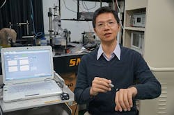
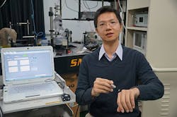
The system, invented by Dr. Sheng-Hao Tseng of NCKU, can be used to quantify the superficial skin absorption and scattering spectrum over a range of wavelengths from 600 to 1000 nm. Specifically, says Tseng, it can detect the concentration of hemoglobin, melanin, collagen, and water of the skin and the condition of melanoma and skin aging, helping doctors diagnose and treat patients faster and more accurately.
Tseng notes that the optical properties of tissues contain very useful information regarding the biological condition and morphological status of tissues. To that end, their optical properties have been used for many diagnostic and therapeutic applications, such as cancer diagnosis, chemotherapy monitoring, photothermolysis, and photodynamic therapy (PDT).
Among many techniques used to determine the optical properties of biological tissues, diffuse reflectance spectroscopy (DRS) has been successfully employed to determine the absorption and scattering properties of deep tissues such as breast tumor, brain, and muscle, according to Tseng.
Tsengâs team has collaborated with the department of dermatology at NCKU to utilize the database to design the âmodified two-layerâ probes to work with the DRS system for skin diagnostic and therapeutic applications.
The diffusing probe with âmodified two-layer geometryâ is a novel design by Tsengâs team. Compared to conventional measurement geometry, the âmodified two-layer geometryâ can be more efficient and accurate in recovering the optical properties of superficial tissues, says Tseng.
In a study using their optical system, findings concluded that collagen concentration, hemoglobin oxygen saturation, and the reduced scattering coefficient of keloids are remarkably different from that of normal skin. The system is also capable of assisting in understanding the functional and structural condition of keloid scars. The research team is currently investigating the utility of the system for other skin-collagen-related studies, notes Tseng.
Full details of the work appear in the Journal of Biomedical Optics; for more information, please visit http://biomedicaloptics.spiedigitallibrary.org/article.aspx?articleid=1351622.
-----
Follow us on Twitter, 'like' us on Facebook, and join our group on LinkedIn
Subscribe now to BioOptics World magazine; it's free!

