FTIR spectroscopy helps predict bone density, fracture risk
Researchers at the University of Eastern Finland (Joensuu and Kuopio, Finland) used bone histomorphometry and Fourier transform infrared (FTIR) spectroscopy to reveal abnormal bone properties in children with vertebral fractures and in children after solid organ transplantation. Bone compositional changes in children with vertebral fractures and after different types of organ transplantation have not been reported previously.
Related: FTIR spectroscopy method characterizes Staph bacteria quickly
Related: IR spectroscopy illuminates protein structure and function
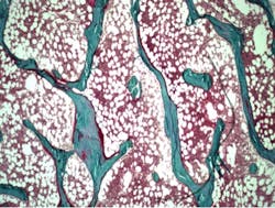
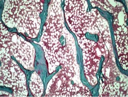
Bone samples were investigated using bone histomorphometry, a microscopic method that provides information about bone metabolism and remodeling. In children with vertebral fractures, there were changes in bone composition, such as lower carbonate-to-phosphate-ratio and increased collagen maturity (determined using FTIR spectroscopy), which could explain the increased fracture risk. The results also suggest that in children who have undergone kidney, liver, or heart transplantation, the various changes related to bone microarchitecture and turnover may be more important predictors of fracture risk than lowered bone mineral density alone. Early detection of such changes in bone quality could help prevent fractures.
Osteoporosis is the most common metabolic bone disease characterized by abnormal bone formation and resorption, which lead to increased risk of bone fractures. However, the present diagnostics based on the measurement of bone mineral density predict fractures only moderately. In addition to decreased bone mineral density, changes in bone quality could explain increased fragility related to osteoporosis. The present study confirmed that bone histomorphometry is needed in clinical practice to study remodeling balance in bone in certain patient groups.
The findings have been published in the Journal of Bone and Mineral Research and the Journal of Bone and Mineral Metabolism.
-----
Follow us on Twitter, 'like' us on Facebook, and join our group on LinkedIn
Subscribe now to BioOptics World magazine; it's free!

