Multispectral imaging method, gold nanotubes destroy cancer cells
Scientists at the University of Leeds in England have shown that gold nanotubes can serve as internal nanoprobes for high-resolution multispectral imaging, as drug delivery vehicles, and as agents for destroying cancer cells. In their study, they demonstrated biomedical utility of gold nanotubes in a mouse model of human cancer.
Related: OCT plays important part in new quantitative imaging biomarker for scleroderma
"High recurrence rates of tumors after surgical removal remain a formidable challenge in cancer therapy," explains Dr. Sunjie Ye, who is based in both the School of Physics and Astronomy and the Leeds Institute for Biomedical and Clinical Sciences at the University of Leeds, and who led the work. "Chemo- or radiotherapy is often given following surgery to prevent this, but these treatments cause serious side effects."
The researchers say that a new technique to control the length of nanotubes underpins the research. By controlling the length, the researchers were able to produce gold nanotubes with the right dimensions to absorb near-infrared (NIR) light—a wavelength for which human tissue is transparent.
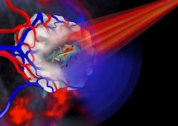
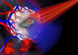
"When the gold nanotubes travel through the body, if light of the right frequency is shone on them, they absorb the light," explains Professor Steve Evans from the School of Physics and Astronomy at the University of Leeds, who was the study's corresponding author. "This light energy is converted to heat, rather like the warmth generated by the Sun on skin. Using a pulsed laser beam, we were able to rapidly raise the temperature in the vicinity of the nanotubes so that it was high enough to destroy cancer cells.”
In cell-based studies, by adjusting the brightness of the laser pulse, the researchers say they were able to control whether the gold nanotubes were in cancer-destruction mode, or ready to image tumors.
To see the gold nanotubes in the body, the researchers used an imaging technique called multispectral optoacoustic tomography (MSOT) to detect the gold nanotubes in mice, in which gold nanotubes had been injected intravenously. It is reportedly the first biomedical application of gold nanotubes within a living organism. It was also shown that gold nanotubes were excreted from the body and therefore are unlikely to cause problems in terms of toxicity, an important consideration when developing nanoparticles for clinical use.
"This is the first demonstration of the production, and use for imaging and cancer therapy, of gold nanotubes that strongly absorb light within the 'optical window' of biological tissue," says study co-author Dr. James McLaughlan from the School of Electronic & Electrical Engineering at the University of Leeds. "The nanotubes can be tumor-targeted and have a central 'hollow' core that can be loaded with a therapeutic payload. This combination of targeting and localized release of a therapeutic agent could, in this age of personalised medicine, be used to identify and treat cancer with minimal toxicity to patients."
The use of gold nanotubes in imaging and other biomedical applications is currently progressing through trial stages towards early clinical studies.
Full details of the work appear in the journal Advanced Functional Materials; for more information, please visit http://dx.doi.org/10.1002/adfm.201404358.
-----
Follow us on Twitter, 'like' us on Facebook, connect with us on Google+, and join our group on LinkedIn
Subscribe now to BioOptics World magazine; it's free!

