Broadband IR light source detects molecular fingerprints of cancer cells with ultrasensitivity
A newly developed broadband and coherent infrared (IR) light source serves as an ultrasensitive detector for the IR molecular fingerprint region, as it can detect minute changes in spectral features from cancer cells or tissue.
Related: Choosing components to image intracellular signals
The Attoscience and Ultrafast Optics Group at the Institute of Photonic Sciences (ICFO; Barcelona, Spain) led the light source's development, in collaboration with the Laboratory for Attosecond Physics at the Max Planck Institute for Quantum Optics (MPQ; Garching, Germany) and Ludwig-Maximilians University (LMU; Munich, Germany).
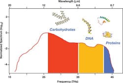
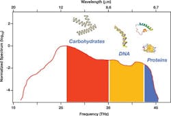
The mid-wave IR (MWIR) is an extremely important range of the electromagnetic spectrum because the wavelength of the light can resonantly excite molecular vibrations. Consequently, shining light through a sample leaves the resonant fingerprints in the spectrum, allowing identification. The absence of light sources that cover enough of the IR spectrum with sufficient brilliance to detect minute concentrations originating from onco-metaboloids has been the main challenge in cancer detection.
The new light source, which addresses this need, exerts extreme control over MWIR laser light with unrivaled peak brilliance and single-shot spectral coverage between 6.8 and 16.4 µm. The emitted radiation is fully coherent and emitted 100 million times per second. Each laser pulse has a duration of 66 fs, which is so short that the electric field oscillates only twice. These characteristics, in combination with its coherence, make the light source a compact and ultrasensitive molecular detector.
Prof. Jens Biegert, who led the work, and his colleagues at ICFO are currently investigating molecular sensitivity for the identification of cancer biomarkers on the single-cell level using all-optical techniques in the MWIR range.
Full details of the work appear in the journal Nature Photonics; for more information, please visit http://dx.doi.org/10.1038/nphoton.2015.179.
Follow us on Twitter, 'like' us on Facebook, connect with us on Google+, and join our group on LinkedIn

