Device couples Raman spectroscopy and microfluidics to detect urinary tract infections faster
Researchers at Jena University Hospital and the Leibniz Institute of Technology (Jena, Germany) have developed a lab-on-a-disc platform that combines Raman spectroscopy and modern microfluidic techniques to cut the time to detect bacterial species that cause urinary tract infections from 24 hours to within 70 minutes.
Related: Optical scanning technique could help diagnose lower urinary tract obstruction
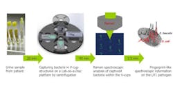
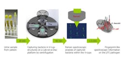
The device is designed to harness centrifugal force to capture the tiny bacteria directly from a urine sample. “Our device works by loading a few microliters of a patient’s urine sample into a tiny chip, which is then rotated with a high angular velocity so that any bacteria is guided by centrifugal force through microfluidic channels to a small chamber where ‘V-cup capture units’ collect it for optical investigation,” explains Ulrich-Christian Schröder, a PhD student at the Jena University Hospital and Leibniz Institute of Technology.
The team's concept then adds Raman spectroscopy to its centrifugal microfluidic platform. “Raman spectroscopy uses the way light interacts with matter to produce ‘unique scattering,’ the equivalent of a molecular fingerprint, which can then be used to identify the types of bacteria present,” says Ute Neugebauer, group leader at the Jena University Hospital and Leibniz Institute of Technology.
In a pilot study, the researchers were able to identify Escherichia coli (E. coli) and Enterococcus faecalis—two species known to cause urinary tract infections—within 70 minutes and directly from patients’ urine samples, Schröder says.
The team envisions general practitioners, using the device to rapidly—while a patient waits—identify the bacteria causing an infection directly within the patient’s bodily fluid so that they can prescribe the appropriate medication and treatment. The next step, says Neugebauer, will involve implementing antibiotic susceptibility testing and automating the sample pre-treatment steps.
Full details of the work appear in the journal Biomicrofluidics; for more information, please visit http://dx.doi.org/10.1063/1.4928070.
Follow us on Twitter, 'like' us on Facebook, connect with us on Google+, and join our group on LinkedIn

