SPECTROSCOPY/ONCOLOGY/GYNECOLOGY: First-ever minimally invasive ovarian cancer screen is spectroscopy-based
Researchers at Northwestern University and NorthShore University HealthSystem (Evanston, IL) have previously demonstrated the ability of partial-wave spectroscopy (PWS) to detect subtle changes in cells that indicate cancer growth in a different area of the body—even when those same cells appear normal under a microscope.1-3 Now, the team has used the same technology to develop the first screening method for early detection of ovarian cancer in humans by examining not the ovaries, but cells brushed easily from the cervix or uterus.4 The work is particularly exciting because currently, there is no reliable way to detect ovarian cancer at early stages.

The outcome of this research could be a minimally invasive detection method using cells collected by a swab, exactly like a Pap smear, explained lead author Dhwanil Damania, who developed the technology. In fact, such a screening could be coupled to the Pap smear. Ovarian cancer ranks fifth in cancer fatalities among American women, and usually goes undetected until it has spread—at which point it is difficult to treat.
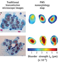
PWS uses light scattering to examine the architecture of cells, and can detect cell features as small as 20 nm along with changes that are the earliest known signs of carcinogenesis. It measures the disorder strength of the cell's nanoscale organization, which is a strong marker for the presence of cancer in the organ or in a nearby organ. Such changes can be seen in cells far from the tumor site or even before a tumor forms. This is the "field effect," a biological phenomenon in which cells located some distance from the malignant or pre-malignant tumor undergo molecular and other changes.
"The changes we have seen in cells have been identical, no matter which organ we are studying," said Vadim Backman, professor of biomedical engineering at Northwestern's McCormick School of Engineering and Applied Science. "We have stumbled upon a universal cell physiology that can help us detect difficult cancers early. If the changes are so universal, they must be very important."
1. H. Subramanian, Proc. Nat. Acad. Sci., 105, 51, 20118–20123 (2008).
2. H. Subramanian, Cancer Res., 69, 13, 5357–5363 (2009).
3. H. K. Roy et al., Cancer Res., 70, 20, 7748–7754 (2010).
4. D. Damania et al., Int. J. Cancer, 1097–0215 (2013).