Dispersive imaging enhances robotic pediatric surgery
JAY ZAKRZEWSKI and KEVIN DIDONA
Raman spectroscopy is experiencing a rapid increase in application integration. While there has been much commercial discussion regarding relatively low-cost, low-resolution, single-point (single-fiber) spectrometers for handheld transportable applications, there has been only limited advancement of higher-performance instruments. Applications that require maximum signal collection and dynamic range, including those that need multichannel or other input-aperture imaging capability, will benefit if the entire height of the tallest spectroscopy CCD can be filled with minimal distortion of the input image.
This requirement is particularly critical in emerging clinical applications such as intraoperative tissue diagnosis. Robotic surgery is revolutionizing cancer treatment in a growing number of hospitals. The Children’s Hospital of Orange County (CHOC; Orange, CA) is one of the leaders in developing this promising technique for pediatric applications. But despite the advances in therapeutic methods such as robotic and laparoscopic surgeries, diagnostic methods simply have not kept pace. This gap led CHOC scientist Chad Lieber and surgeon Mustafa Kabeer to undertake the development of Raman spectroscopy for intraoperative pediatric-tissue diagnosis.
While Raman spectroscopy has been demonstrated in various medical diagnostic applications, almost none have entailed pediatric patients and their respective diseases. Furthermore, the few in vivo reports have generally targeted readily accessible anatomic locations such as skin and cervix. To obtain Raman spectra from internal tissues, the robotic approach provides an ideally suited vehicle. Once inside the body, the collection of relevant tissue Raman spectra in clinically feasible times necessitates maximization of optical throughput.
The CHOC researchers have developed a portable Raman system for their study that utilizes custom fiber probes and a Headwall Raman Explorer spectrograph. The Raman Explorer is an aberration-corrected, high-reciprocal-dispersion, retroreflective, concentric-imaging spectrograph with multiple inputs and high efficiency through resonance, which produces minimal image blur over the full CCD focal plane with exceptionally high signal-to-noise efficiency and f/2.4 throughput. This design images 1:1 straight slit or stacked linear arrays of optical fibers covering the full height of spectroscopy CCDs with optimal channel separation and no image curvature.
Lieber and Kabeer’s selection of the Raman Explorer was based on comparative studies with transmissive spectrographs, which showed the throughput and dynamic range of the Raman Explorer to be well suited to the turbid, bloody internal environment of the human body.1 Although their current research is in the early stages, the CHOC team aims to provide the first reported use of Raman spectroscopy for characterization of a number of pediatric cancers during open, laparoscopic, and robotic surgeries.
Spectral and spatial resolution
Most commercially available dispersive-spectrometer optical designs have difficulty imaging all field points of the full Raman spectral bandwidth onto a flat focal plane simultaneously. Czerny-Turner designs are typically throughput-limited to approximately f/4 or slower, and exhibit relatively high image distortions as one moves farther away from the central input axis (spatially over the slit height) as well as along the spectral axis. Torroidal mirrors are often used to minimize this effect, although this method provides optimized correction for only one point on the focal plane. Others, based on axial-transmissive designs, suffer from chromatic aberrations, smile, and keystone at short focal lengths. For best results, these often require the use of curved input apertures, nonstandard refractive lens material selection, and wavelength optimized antireflective coatings on each lens surface to compensate for chromatic aberrations, reflection losses, and potential ghost images. In addition, common axial-transmissive designs suffer from throughput vignetting at both the short and long wavelength regions.
Any degradation of an image that is launched into a spectrograph—whether via a single fiber core, a large stack of multiple fibers, or an input slit—will impact the instrument’s spectral resolving power, optical-throughput efficiency, and multichannel crosstalk performance. The quality of both spectral and spatial input-image reproduction at the spectrograph exit focal plane is often overlooked when optimizing spectrometer designs.
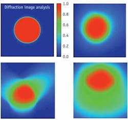
We evaluated the spectral and spatial imaging performance of the Raman Explorer to obtain spectral intensity data that can be interrogated to display image shape, a comparison of FWHM spectral and spatial resolution resulting from 50-µm-diameter fiber cores, with and without a 10-µm-slit aperture, and quality of horizontal and vertical image maintenance (keystone and smile distortion eliminated). The ray-trace images provide examples of nominal spectral image distortion (see Fig. 1). A theoretical 50-µm-diameter image of uniform intensity at a specific wavelength is shown at Fig. 1, top left. At various spatial positions over a focal plane, one would like to maintain this shape or the shape in the image at top right, as much as possible. If a spectrograph design prescription is not optimized, aberrations distort the size and shape of the spectral image, causing rays intended for that wavelength and spatial position to fall on adjacent locations and thus corrupting the spectral purity of the measured results.
Examples of emerging image distortions are shown in Fig. 1 bottom, left and right. The distortions are minimal in relation to the extent they can degrade in commonly accepted research-grade spectrograph designs. The data demonstrate the Raman Explorer does an exceptional job of maintaining high spatial and spectral integrity (similar to top right) over the entire focal plane.
Neon and argon illumination
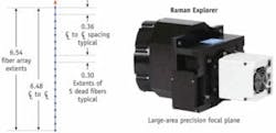
We also evaluated fine spectral and spatial imaging resolution capabilities and signal throughput characteristics of the Raman Explorer 785 f/2.4 spectrograph over the area of an Andor Newton camera that included a 2048 × 512 array with 13.5 µm square pixels. To enable imaging of narrow spectral lines, we aligned a test fiber assembly to the channel 1 entrance aperture of a Raman Explorer 785. The test fiber assembly comprises an array of 19 live 50/60/70 µm fibers, each separated by five dead fibers (see Fig. 2). This fiber assembly allows the placement of live 50-µm-core fiber images over 6.5 mm of the available 7 mm of entrance aperture height at 360 µm center-to-center distances.
Using neon and argon atomic line sources to illuminate spectral lines across the full spectral bandwidth of the CCD, Headwall engineers used the following method to capture meaninful spatial and spectral data points: focus the fiber images and adjust the light-source intensity and camera integration time to just reach, but stay within, saturation limits of brightest line; set the integration time so that readout smear between channels was minimized, set background subtraction, and captured and stored the image; same conditions as before, except with a 10 µm slit placed over the ferrule face; take and store image.
Spectral images were produced by neon and argon illumination through the Raman Explorer 785, in which the fiber is 50 µm in diameter and the pixel size is 24 µm square (see Fig. 3). Horizontal, vertical, and FWHM imaging was pixel-limited. Note, the pixels are 13.5 µm square, the fiber is 50 µm core with a 10 µm cladding, and in all cases the images are maintained with superb accuracy.
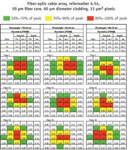
The Raman Explorer, designed for 785 nm laser stimulation, and integrating a CCD with a 13.5 µm pixel pitch and 10-µm-wide entrance aperture, is capable of 3.7 cm–1 spectral resolution over the fingerprint region, and 2.6 cm–1 over the CH stretch region.
The spectral and spatial imaging properties of a spectrograph play an important role in the ability of a spectrometer to produce spectrally pure results, a particularly critical requirement in intraoperative tissue imaging. The Raman Explorer spectrographs produce minimal image blur and no smile or keystone over the full CCD focal plane, which enables higher signal levels, lower optical noise, and high spectral resolution over the full Raman bandwidth.
REFERENCE
- C.A. Lieber, E.M. Kanter, and A. Mahadevan-Jansen, Applied Spectroscopy., in press.
Jay Zakrzewski is director of business development and Kevin Didona is senior design engineer at Headwall Photonics (Fitchburg, MA). Contact Zakrzewski at [email protected].