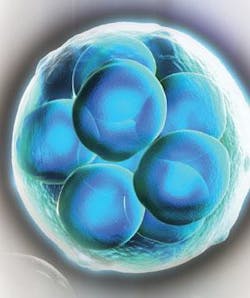
NIR spectroscopy enables embryo assessment
PIETER ROOS
Because biospectroscopy can produce a complete profile of a biological sample’s metabolic activity, it solves a longstanding problem in in vitro fertilization.
The American Society for Reproductive Medicine agrees with in vitro fertizilation (IVF) practitioners, patients, and insurance providers on a key goal: reducing multiple gestations without compromising pregnancy rates. Noninvasive assessment of gamete and embryo reproductive potential has long been the “holy grail” of IVF. Poor embryo quality has been linked to both morphological abnormalities and metabolic errors. The availability of embryo metabolomic profiling would lead to markedly improved treatment outcomes and success rates.
Metabolomics is a form of systems biology that provides a biochemical “snapshot” of the small molecule inventory produced during cellular metabolism that effectively reveals the physiological status of an organism. Metabolomic profiling would empower practitioners to more confidently move from multiple embryo transfer to single embryo transfer (SET) as the new paradigm of clinical practice in IVF. Medical experts agree that metabolomic testing is likely to result in the following benefits:
- enhanced treatment outcomes, and a reduction in the multiple birth rate;
- reduced number of IVF treatment cycles needed to achieve a healthy pregnancy;
- a reduction in medical risk to mother and babies from multiple gestations, resulting in reduced medical costs;
- reduction in the time and cost (both financial as well as emotional) of achieving a successful pregnancy;
- enhanced quality of donor egg and sperm banks;
- expansion of the IVF market as more couples can afford to seek treatment for infertility; and
- broader insurance coverage for IVF procedures.
During embryo culture, and at the time of embryo selection prior to transfer, a portion of the liquid culture media that bathes the embryo can be sampled and analyzed (see figure). Molecular Biometrics (Chester, NJ) has developed a noninvasive testing system, ViaMetrics-E, to do just that—and thus provide IVF laboratories with embryo viability evaluation capabilities.
The result is an assessment of reproductive potential, or viability, for each individual embryo based on the levels of various nutrients and metabolic by-products present in the media sample after the culturing period. All culture samples used in the metabolomic profiling are excess specimens that are either discarded or stored after IVF; only small sample volumes (7 μl) are required for analysis. Proprietary bioinformatics technology captures and analyzes multiple biomarker data, and produces results in about one minute.
Instrumentation
Biospectroscopy is an optical-based analytical technology that provides a simultaneous and integrated measure of all metabolic activities taking place in a biological sample. Each resulting spectrum translates into a unique “fingerprint” or “metabolomic profile” that defines the metabolic status of the target sample and thus of the biological host from which the sample was taken.
Multiple characterization tools, both chromatographic and spectroscopic, assist the process of metabolomics. Molecular Biometrics’ technology platform (see “How ViaMetrics works”) uses capillary electrophoresis (CE), mass spectrometry (MS), and Raman and near-infrared (NIR) spectroscopy. But NIR spectroscopy is the primary instrumentation candidate.
NIR, the region of light immediately adjacent to the visible range, falls between 750 and 3000 nm in wavelength. According to the principles of quantum physics, molecules can only assume discrete energy levels. Similar to the vibrating string of a musical instrument, the vibration of a molecule has a fundamental frequency, or wavelength, as well as a series of overtones. The unique spectrum shape for any material is the result of the absorbance of the characteristic fundamentals and overtones. NIR spectra result from a harmonic overtone and combination bands caused by vibrations of O-H, C-H, N-H and S-H groups.
NIR spectroscopic analysis is simple: expose a target sample, such as a biological fluid, to a range of light wavelengths, and measure the characteristic absorbance spectrum of the sample. Because the molecular structure of most compounds is very complex, the resulting spectra are actually the result of many overlapping peaks. In addition, the amount of NIR light absorbed is directly proportional to the quantity of molecules present in the sample. Generally speaking, people performing NIR analysis must then identify and characterize specific features in the spectra by means of statistical methods. Chemometrics and bioinformatic software is designed to accomplish this task.
The technology can be easily compacted into a portable device that requires little maintenance, and is relatively inexpensive.Metabolomics vs. morphology
Four proof-of-principle studies have been published comparing the results of morphology—the parameter traditionally used for embryo assessment—versus metabolomics on the ViaMetrics-E platform (Seli et al., Fertil Steril 2007; Scott et al., Fertil Steril 2008; Vergouw et al., Human Reprod. 2008; Nagy et al., RBM online). In every case, the studies show that ViaMetrics-E produces greater accuracy than morphology in assessing single embryo transfer (SET; see table). The table summarizes the four studies. Oocytes are fertilized and the resulting embryos are grown for up to five days; the day of transfer (in this case, day two) refers to when they are placed back into the patient.
Pieter Roos, Ph.D., is associate director of R&D at Molecular Biometrics, 5 Topping Way, Chester, NJ 07930; www.molecularbiometrics.com. Contact him at [email protected].
How ViaMetrics works
At the heart of the ViaMetrics-E system is a compact spectrograph manufactured by B&W Tek (Newark, DE; see figure). The spectrograph uses an InGaAs array with 512 pixels manufactured by Goodrich Sensors Unlimited (Princeton, NJ). The array is thermoelectrically cooled for improved sensitivity and signal stability, and provides fast scan capabilities up to 3 ms per spectrum. The disposable sample cell holder (patent pending) requires very small volumes (7 µl) of the spent media in which the embryo was cultivated. The sample is illuminated by a very stable 5 W tungsten light source (also by B&W Tek). The data is collected and processed by a panel PC with a 7 in. touch-screen interface running proprietary software.Through the use of a highly sensitive method of biomarker identification, analysis can be performed on-site in just minutes. The method provides increased accuracy in the assessment of viability without compromising the embryo, helping guide treatment options for patients undergoing IVF. The system is a small, user friendly instrument for the embryology lab that offers a noninvasive assessment of embryo viability with complete analysis in less than one minute. It requires only 7 µL of spent culture media. The system also provides a seamless software interface for medical records and data mining.


