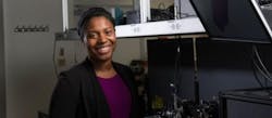
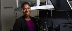
Medical imaging innovator Hendon wins Presidential Early Career Award
Christine Hendon, assistant professor of electrical engineering at Columbia Engineering (New York, NY), has won the Presidential Early Career Award (PECASE), the highest honor the U.S. government gives to young scientists and engineers. Hendon, who develops innovative medical imaging instruments for use in surgery and breast cancer detection, is one of 102 researchers named by President Obama on January 9, 2017.
Related: Photonics is key for Cancer Moonshot
Hendon is developing optical imaging and spectroscopy instruments for surgical guidance, aiming to provide surgeons with a clear understanding of the tissue on which they are operating. She uses near-infrared spectroscopy and optical coherence tomography (OCT), which provides depth-resolved, high-resolution images of tissue microstructure in real time and noninvasively. Using OCT, a surgeon could image a wide area of tissue and, unlike invasive biopsies, remove as little tissue as possible.
Hendon is currently working with Vivek Iyer, a cardiac electrophysiologist at Columbia University Medical Center, to explore the use of OCT and spectroscopy in the treatment of heart arrhythmias, where surgeons often use a catheter to detect abnormal electrical signals and then apply radiofrequency energy to remove scar tissue in the malfunctioning area.
Other projects running in Hendon's Structure Function Imaging Laboratory include using optical tools to detect and image breast cancer. She is working with breast surgeon Sheldon Feldman and pathologist Hanina Hibshoosh at Columbia University Medical Center to identify tumors localized to the duct and eventually to image lesions over time to determine which are likely to progress to cancer. Hendon is also collaborating with Columbia Engineering associate professor Kristin Myers on using imaging to assess the mechanical properties of the cervix in relation to preterm birth.
For more information on Hendon's work, please visit https://structurefunctionlab.com.

