Doppler OCT imaging sheds light on health of baby's heart
For the first time, researchers at Case Western Reserve University (Cleveland, OH), using an optical coherence tomography (OCT) method, have been able to visualize in 3D the stresses induced by flowing blood in an embryonic heart. The method is promising for learning how and why heart defects develop.
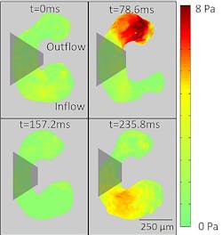
The researchers, led by Andrew M. Rollins, an associate professor of biomedical engineering at Case Western Reserve University, looked at a particular type of stress in the heart known as shear stress, which is simply the parallel force of one material sliding along another. In the developing heart, shear stress is induced in the heartâs own endocardial cells as blood cells rush past them. Normally, such shear stress helps to control and regulate cellular processes involved in heart development. Even tiny aberrations in the heart beat, however, can alter blood flow patterns and change these developmental forces, leading to congenital heart defects such as abnormal valve formation.
âAll previous attempts at shear-stress mapping have been two-dimensional, but the 3D geometry of the embryonic heart is changing hour by hour at these early stages, and the shape of the heart twists and turns as it develops,â says Rollins, âso a 2D projection doesnât really provide a good approximation.â
Rollins and colleagues used Doppler OCT, which shines a beam of infrared (IR) light on tissue. Then, the reflections of that light produced at varying depths are used to make a high-resolution image. The technique has been used to image the interior of blood vessels and is routinely used by ophthalmologists to examine the retina.
âWe can use this technique to figure out just how function or dysfunction fits into normal heart development and the development of heart defects,â Rollins adds. âAn understanding of normal and abnormal development is critical for preventing and treating these defects.â
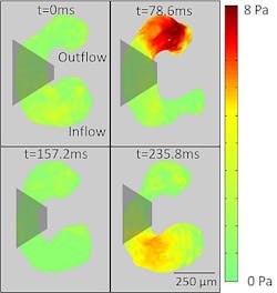
In laboratory experiments, Rollins and colleagues directly measured the heart structure and blood flow within the developing hearts of quail embryos. The data was then used to create 4D images, which showed that âlocations of high shear correspond with locations of future valve formation,â he says. âNow we are investigating the effects of abnormal shear caused by alcohol exposure on valve development.â
The researchers hope to take what they have learned from these preliminary animal tests and develop a way to apply this method to humans. The ultimate goal is to develop a tool that doctors can use to decide if early intervention could put a developing heart back on the right track, preventing a defect.
The work appears in Biomedical Optics Express; for more information, please visit http://www.opticsinfobase.org/boe/abstract.cfm?uri=boe-3-11-3022.
-----
Follow us on Twitter, 'like' us on Facebook, and join our group on LinkedIn
Laser Focus World has gone mobile: Get all of the mobile-friendly options here.
Subscribe now to BioOptics World magazine; it's free!