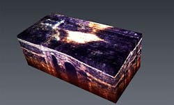
Spectroscopic OCT technique reveals true colors of living tissue
| A spectroscopic optical coherence tomography (OCT) technique developed by Duke University researchers enabled them to image cellular reactions beneath the skinâs surface in true color. (Image courtesy of Adam Wax) |
Researchers at Duke University (Durham, NC) have developed a new technique that detects and shows in vivid shades of red the levels of hemoglobin being carried by blood vessels, even the tiniest ones whose diameter can be as narrow as a few red blood cells. Other molecules can also be visualized, including medical dyes introduced to trace important biological mechanisms. The technique could have significant implications both clinically and in basic science laboratories by providing new perspectives of basic biological functions.
To achieve this colorful view below the skin, the researchers used a modified optical coherence tomography (OCT) technique to provide cross-sectional images of biomolecules. Conventional OCT allowed the team to see structures such as blood vessels and even capillaries but it didnât provide functional information such as absorption, which also gives the true color of the structures, says Francisco Robles, a graduate student at Dukeâs Pratt School of Engineering.
Adam Wax, Theodore Kennedy associate professor of biomedical engineering, says that the team expects that the new technique will have several important applications, such as visualizing tumor development processes like angiogenesis and oxygen deprivation. It also could help in detecting disease of the eye, especially those that impact the tiny vessels of the eye, as well as monitoring drug delivery and effectiveness, he says.
To achieve this ability to see true colors, Robles developed a novel method for collecting and interpreting the information provided by the OCT procedure to simultaneously include information regarding the wavelengths or colors of light. Each point on the collected 3-D image contains a great deal of useful information that is useful in reconstructing whatâs happening at the molecular level, says Robles. The data not only helps to create 3-D images but it also contains important spectral information, he adds.
The hemoglobin being carried by red blood cells provides the absorption that makes the process work. In a sense, the hemoglobin, which carries oxygen, acts as a ânaturalâ contrast agent because of its absorption properties, which give blood its red color. Many disorders can be characterized by the levels of blood oxygen, a characteristic that can be detected in true shades of red by the new technique.
The team tested the new system in living mice. When conventional OCT was employed, certain structuresâsuch as muscle and vesselsâcould be observed. However, these images were black and white and couldnât reveal information about the tissue function.
âWith the new system, we observed a wealth of information we couldnât before,â says Robles. âThe muscle layer at the surface was relatively colorless because of low hemoglobin levels. However, as the light continued through the blood vessels below, a red shift in color was clear. To our knowledge, these are the first micron-scale cross-sectional images of living tissue in true color.â
The National Institutes of Health supported this work, which has been published online by Nature Photonics. For more information, please visit http://www.nature.com/nphoton/journal/vaop/ncurrent/full/nphoton.2011.257.html.
-----
Follow us on Twitter, 'like' us on Facebook, and join our group on LinkedIn
Follow OptoIQ on your iPhone; download the free app here.
Subscribe now to BioOptics World magazine; it's free!
