Newly achieved meter-scale OCT expands its utility in medicine
Researchers at the Massachusetts Institute of Technology (MIT; Cambridge, MA), in collaboration with Praevium Research (Santa Barbara, CA), Thorlabs (Newton, NJ), and Acacia Communications (Maynard, MA), have achieved optical coherence tomography (OCT) images of cubic meter volumes. With OCT's ability to provide information on material composition, subsurface structure, coatings, surface roughness, and other properties, this advance could open up many new uses for OCT in medicine, among other applications. The achievement also represents important progress toward developing a high-speed, low-cost OCT system on a single integrated circuit (IC) chip.
Related: Improved OCT imaging with VCSEL technology
The research team's study demonstrates OCT with at least an order-of-magnitude larger depth range and volume compared to previous demonstrations of three-dimensional (3D) OCT, according to James G. Fujimoto of MIT, whose group and collaborators first invented OCT in the early 1990s. The imaging method is now the standard of care in ophthalmology, and is increasingly being used in cardiology and gastroenterology.
In a paper describing the work, the researchers report high-speed, 3D OCT imaging with 15 µm resolution over a 1.5 m area. They demonstrated the new OCT approach by imaging a mannequin, a bicycle, and models of a human brain and skull. They also conducted measurements of objects ranging in scale from meters to microns.
In addition to the advantages of high speeds and fine resolution, OCT enables imaging, profiling, and distance measurement at multiple depths simultaneously while rejecting stray light. "Long-range OCT is a new range of operation that requires extremely high-performance light sources, integrated optical receivers, and signal processing," Fujimoto explains. Range in OCT refers to the depth range over which measurements can be simultaneously taken. It is possible to position the center of the OCT range very close to or far away from the imaging instrument.
The new method could enhance medical imaging, for example, by providing 3D measurements in laparoscopy or mapping structures such as the upper airway.
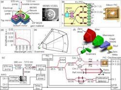
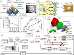
The light source that enables meter-range OCT is a tunable vertical cavity surface-emitting laser (VCSEL) developed by Thorlabs and Praevium Research. It uses a MEMS device to rapidly change, or sweep, the laser's wavelength over time to perform what is called swept-source OCT. "Research by our group at MIT and our collaborators at Praevium Research and Thorlabs indicated that the coherence length of the VCSEL source was orders of magnitude longer than other swept-laser technologies suitable for OCT, which suggested the possibility of long-range OCT imaging," says Ben Potsaid of MIT and Thorlabs, and a coauthor of the paper.
Although the researchers have experimented with the VCSEL light source for several years, light detection and data acquisition remained a challenge. These hurdles were overcome by advanced optical components designed for telecommunications applications. In their new work, the researchers used a new silicon photonics coherent optical receiver developed by Acacia Communications that replaced several bulky OCT components with integrated optics on a tiny, low-cost, single-chip photonic integrated circuit (PIC). Importantly, the PIC receiver supports the very high electrical frequencies and wide range of optical wavelengths required for swept-source OCT while also enabling what is known as quadrature detection, which doubles the OCT imaging range for a given data acquisition speed.
In the paper, the researchers showed that meter-range OCT can obtain a strong signal from surfaces of varying geometry and materials. Their tests also indicated the technique's performance has not reached the fundamental limits for the VCSEL laser source or PIC receiver. They are now working to develop and utilize even more low-cost, high-speed components, with the goal of speeding up the data acquisition and processing steps. This could eventually allow real-time OCT imaging using customized IC chips.
"As PIC technology continues to advance, one can realistically expect full OCT systems on a single chip within the next five years, dramatically lowering the size and cost," says Chris Doerr of Acacia Communications, a coauthor of the paper. "This would allow more people all over the world to benefit from OCT and open up new applications."
The researchers note that the method could also have utility in industrial and manufacturing settings, where it could potentially be used to monitor processes, take technical measurements, and nondestructively evaluate materials.
Full details of the work appear in the journal Optica; for more information, please visit https://doi.org/10.1364/optica.3.001496.

