Eye test that pairs two in vivo imaging methods may detect Parkinson's earlier

Researchers at the University College London (UCL; England) Institute of Ophthalmology have demonstrated a new low-cost, noninvasive eye test that incorporates optical coherence tomography (OCT) to help detect Parkinson's disease before symptoms like tremors and muscle stiffness develop.
Related: Laser-based eye test for early detection of Alzheimer's
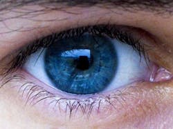
The test, which uses OCT and longitudinal in vivo imaging with detection of apoptosing retinal cells (DARC), can observe changes in the retina that can be seen in Parkinson's before changes in the brain occur and the first symptoms become evident. Using ophthalmic instruments that are routinely used in optometrists and eye clinics, the scientists were able to use the new imaging technique to observe these retinal changes at an early stage.
In addition to allowing earlier diagnosis of Parkinson's, the test could also be used to monitor how patients respond to treatment. The technique has already been tested in humans for glaucoma and trials are due to start soon for Alzheimer's disease.
Following the observation of retinal changes in the experimental model, Professor Francesca Cordeiro, UCL Professor of Glaucoma & Retinal Neurodegeneration Studies, and her team treated the animals with a newly formulated version of the anti-diabetic drug Rosiglitazone, which helps to protect nerve cells. After using this drug, there was clear evidence of reduced retina cell death as well as a protective effect on the brain, which suggests that it could have potential as a treatment for Parkinson's disease.
The technology is being patented by UCL's research commercialization company UCL Business.
Full details of the work appear in the journal Acta Neuropathologica Communications; for more information, please visit http://dx.doi.org/10.1186/s40478-016-0346-z.