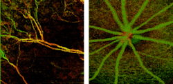
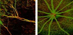
Wasatch Photonics unveils OCT angiography technique
Gratings, spectrometers, and optical coherence tomography (OCT) instrumentation developer and maker Wasatch Photonics (West Lafayette, IN; Logan, UT; and Durham, NC) has launched its WP MicroAngio imaging device that delivers high-resolution angiographic imaging for research and original equipment manufacturer (OEM) applications.
Related: OCT angiography: A new approach with 'gold standard' capabilities and more
MicroAngio technology allows visualization of blood vessels at a microscopic level without using external contrast agents. The device, based on a variation of OCT, creates a three-dimensional (3D) profile of the microvasculature. WP MicroAngio has utility in several medical research applications related to animal model imaging in ophthalmology, dermatology, and oncology, among others.
Unlike techniques like fluorescence and x-ray fluoroscopy, MicroAngio does not require the injection of external contrast agents that can interfere with the physiology of the sample. In addition, the technique provides 3D localized information compared to the 2D data obtained from most other existing techniques.
OCT angiography techniques are already finding use in clinical ophthalmology. MicroAngio is expected to provide the same capability for animal research applications in ophthalmology and extend it to other medical research applications.
Nishant Mohan, director of Product Management and Marketing, systems division at Wasatch Photonics, explains that MicroAngio comes with customized probes and fixtures for small animal retinal, dorsal window chamber, and brain imaging, as well as a suite of algorithms for data analysis essential for angiographic imaging.
MicroAngio's OCT angiography software allows selection of processing based on only intensity or phase of the signal, or a combination of both. The system will also provide techniques to allow for motion correction that is essential for artifact-free imaging.
For more information, please visit http://wasatchphotonics.com.

