PRIMARY CARE SCREENING/OCT/DISEASE DETECTION: Handheld OCT device promises screening for retinal disease in primary care

A new optical coherence tomography (OCT) device about the size of a handheld video camera could enable primary care physicians to detect retinal diseases—including diabetic retinopathy, glaucoma, and macular degeneration—in their early stages to improve treatment outcomes. Researchers at the Massachusetts Institute of Technology (MIT; Cambridge, MA) say that while others have created handheld devices using similar technology, this is the first to combine such cutting-edge technologies as ultra-high-speed 3D imaging, a tiny microelectromechanical systems (MEMS) mirror for scanning, and a technique to correct for motion artifacts. These innovations, they explain, should allow comprehensive data collection with just one measurement.
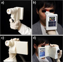
Normally, retinal disease is diagnosed by an ophthalmologist or optometrist using tabletop instruments. But few people visit such specialists regularly, so the MIT group, in collaboration with the University of Erlangen (Nuremberg, Germany) and Praevium/Thorlabs, developed the portable instrument to improve public access to eye care. Screening technology is important because many eye diseases could be detected and treated long before symptoms arise. "Handheld instruments can enable screening a wider population outside the traditional points of care," said MIT's James Fujimoto, an author on the paper describing the work.1
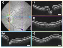
Using a MEMS mirror to scan the imaging beam enabled miniaturization of the hardware. Making multiple high-speed 3D scans of the same area from different directions compensates for instability inherent in a handheld instrument. The researchers' tests showed that the device can produce images comparable in quality to conventional OCT instruments.
The next step is to evaluate the technology in a clinical setting and then find a way to support or lower its cost. Fujimoto thinks that handheld OCT technology may one day enhance other medical specialties, such as surgical guidance and military medicine.
1. C. D. Lu et al., Biomed. Opt. Exp., 5, 1, 293–311 (2013).