Laser endomicroscopy method could enable early pancreatic cancer detection
Knowing that patients typically do not show symptoms of pancreatic cancer until it is advanced, making early diagnosis and treatment a challenge, a team of researchers at The Ohio State University Wexner Medical Center (Columbus, OH) used a technique called endoscopic ultrasound (EUS)-guided needle-based confocal laser endomicroscopy (nCLE) to definitively diagnose cysts in the pancreas with unprecedented accuracy.
The current standard involves testing the fluid inside the cysts. It correctly identifies them as benign or precancerous 71% of the time. Researchers found that when the virtual biopsy is added to the standard of care, the diagnostic accuracy jumps to 97%.
"Pancreatic cysts are common, and it can be difficult to distinguish the benign cysts from those destined to become cancerous, but this procedure allows us to do that quickly and with confidence," says Dr. Somashekar Krishna, a gastroenterologist and lead author of the study. "We hope that, at the end of the day, we are saving lives either by diagnosing pancreatic cancer early on before it develops into cancer, or we are preventing unnecessary surgery of a benign, harmless pancreatic cyst."
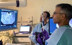
The EUS-nCLE method tested in the researchers' study provides doctors with a microscopic view of the cyst wall, which is produced by a tiny scope that emits laser light inside the cyst. This allows doctors to determine almost immediately if it is precancerous.
"Many times, we are able to tell the patient right after the procedure, 'You have a precancerous cyst, and we need to send you to the surgeon to have it removed,'" says Krishna, who is an associate professor in Ohio State's College of Medicine and is also affiliated with The Ohio State University Comprehensive Cancer Center - Arthur G. James Cancer Hospital and Richard J. Solove Research Institute (OSUCCC - James).A majority of patients get diagnosed with pancreatic cysts incidentally when getting a MRI or CT scan for another reason. Nearly 40% of MRIs done of the abdomen reveal pancreatic cysts and the chance of having them increases with age.
The researchers are now working to train doctors at hospitals nationwide to perform the diagnostic method and read the images provided by the scope to catch dangerous cysts and prevent pancreatic cancer for more patients. They're also working to develop artificial intelligence that will flag cases that are likely precancerous so doctors can act quickly.
Full details of the work appear in the journal Clinical Gastroenterology and Hepatology.
Source: The Ohio State University Wexner Medical Center press release
Got biophotonics-related news to share with us? Contact Lee Dubay, Associate Editor, BioOptics World
Get even more news like this delivered right to your inbox

