Quantum dot approach helps increase resolution in optical microscopy
Recognizing the considerable effort to overcome the diffraction limit in optical microscopy and therefore increase its resolution, a team of researchers at Julius Maximilian University of Würzburg and the Technical University Dresden (TU Dresden; both in Germany) has shown that it is possible to measure near-fields with significantly less effort. To do so, the team used a biomolecular transport system with quantum dots (small fluorescent particles that measure only a few nanometers) to slide many optical nanoprobes over a surface.
Related: Nanoworms with shining quantum dots for a new dimension in microscopy
The system involves motor proteins and microtubules to make the quantum dots pass over the object to be examined. "These two elements are among the fundamental components of an intracellular transport system," explains Stefan Diez, Chairholder of BioNanoTools at the B CUBE - Center for Molecular Bioengineering at TU Dresden. "Microtubules are tubular protein complexes, up to several tenths of millimeters long, that form a major network of transport routes inside cells. Motor proteins run along these routes, transporting intracellular loads from one place to another."
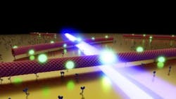
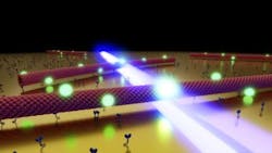
The research team took advantage of this concept, but in reverse order: "The motor proteins are fixed to the sample surface and pass the microtubules over them—a kind of 'stagediving' with biomolecules," says Heiko Gross, a PhD student in Bert Hecht's research group at JMU. The quantum dots serving as optical probes are attached to the microtubules and move together with their carrier.
Since a single quantum dot would take a very long time to scan a large surface area, the researchers used large amounts of quantum dots and motor proteins that move at the same time to scan a large area in a short time. "Using this principle, we can measure local light fields over a large area with a resolution of <5 nm using a setup that resembles a classical optical microscope," Gross explains.
The physicists tested their method on a thin layer of gold with narrow slits <250 nm wide. The slots were illuminated from below with blue laser light. "Light passing through these narrow gaps is limited to the gap width, making it ideal for demonstrating high-resolution optical microscopy," Gross says.
During the measurement, a swarm of microtubules simultaneously glides in different directions across the surface of the gold layer. Using a camera, the position of each transported quantum dot can be exactly determined at defined time intervals. If a quantum dot now moves through the optical near-field of a slit, it lights up more strongly and therefore acts as an optical sensor. Since the diameter of the quantum dot is only a few nanometers, the light distribution within the slot can be determined with extreme precision, thus circumventing the diffraction limit.
Another nice feature of this novel approach is that because of its length and strength, a microtubule can move in an extremely straight and predictable fashion across the motor-coated sample surface. "This makes it possible to determine the position of the quantum dots 10X more accurately than with previously established high-resolution microscopy methods," explains Jens Ehrig, former postdoctoral fellow in the Diez group and current head of the Molecular Imaging and Manipulation facility at the Center for Molecular and Cellular Bioengineering (CMCB) at TU Dresden. Furthermore, it excludes disturbances caused by artifacts because of near-field coupling. Since the transport system consists of only a few molecules, its influence on the optical near-fields is negligible.
The researchers hope to use their idea to establish a new technology in the field of surface microscopy. In a next step, they want to use the molecular transport system to couple quantum dots to specifically prepared optical near-field resonators to study their interaction.
Full details of the work appear in the journal Nature Nanotechnology.

