Deep learning method could improve optical microscopy
A team of researchers at the University of California Los Angeles (UCLA) used a form of machine learning called deep learning, which uses multi-layered artificial neural networks to automate data analysis and reconstruct a hologram to form a microscopic image of an object, thereby improving optical microscopy.
Related: Holographic microscope tracks sperm motion in 3D
The holographic imaging technique produces better images than current methods that use multiple holograms, the researchers say, and it is easier to implement because it requires fewer measurements and performs computations faster.
The research was led by Aydogan Ozcan, an associate director of the UCLA California NanoSystems Institute and the Chancellor's Professor of Electrical and Computer Engineering at the UCLA Henry Samueli School of Engineering and Applied Science; and by postdoctoral scholar Yair Rivenson and graduate student Yibo Zhang, both of UCLA's electrical and computer engineering department.

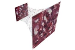
In one study, the researchers produced holograms of Pap smears, which are used to screen for cervical cancer, as well as blood and breast tissue samples. In each case, the neural network learned to extract and separate the features of the true image of the object from undesired light interference and from other physical byproducts of the image reconstruction process.
"These results are broadly applicable to any phase recovery and holographic imaging problem, and this deep-learning-based framework opens up myriad opportunities to design fundamentally new coherent imaging systems, spanning different parts of the electromagnetic spectrum, including visible wavelengths and even x-rays," Ozcan says.
Another advantage of the new approach was that it was achieved without any modeling of light-matter interaction or a solution of the wave equation, which can be challenging and time-consuming to model and calculate for each individual sample and form of light.
In a second study, the researchers used the same deep-learning framework to improve the resolution and quality of optical microscopic images.
That advance could help diagnosticians or pathologists looking for very small-scale abnormalities in a large blood or tissue sample, and Ozcan says it represents the powerful opportunities for deep learning to improve optical microscopy for medical diagnostics and other fields in engineering and the sciences.
Full details of the work appear in the journals Light: Science and Applications and Optica, respectively.